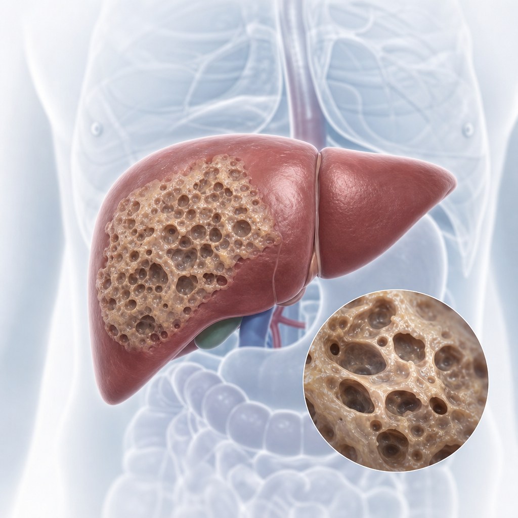
Alveococcosis treatment at Visus Medical without surgery
For residents of Kyrgyzstan, we adapt the protocol to local realities — from logistics to repeat test access. We advise patients from Kyrgyzstan to track their progress systematically. Alveococcosis is not an “ordinary cyst.” The parasite grows infiltratively, invading liver tissue, and often mimics a malignant tumor. We do not promise miracles to everyone: after reviewing your CT/MRI we say honestly whether a non-surgical protocol fits your case.
The Visus Medical proprietary method aims to suppress parasite viability and stabilize or regress the lesion under imaging control. The course is outpatient — no general anesthesia and no liver resection when the clinical situation allows.
Patients come from Russia, Kazakhstan, Kyrgyzstan, and Tajikistan — often after being offered major surgery or refused it. Evidence from Kyrgyzstan shows early therapy yields better outcomes. Many start with a remote review of their scans.
- Focus specifically on the alveolar form (AE), not cystic echinococcosis.
- CT/MRI review before the visit — free initial assessment.
- Outpatient care without hospitalization when clinically justified.
- We work with recurrences and inoperable cases when indicated.
- Dynamic imaging follow-up at every stage.
- Coordination with local physicians in the patient’s country of residence.
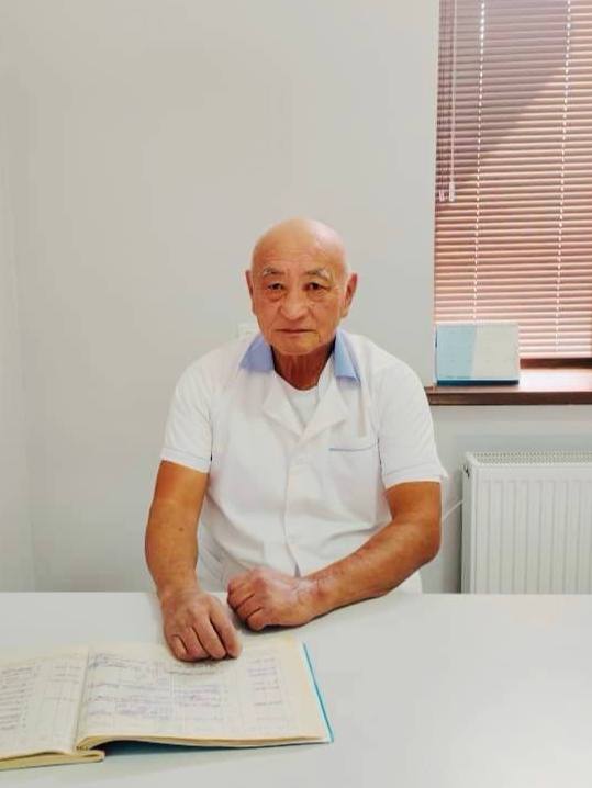
Elfréntiy Li
Chief physician — parasitologist, traditional medicine specialist
- 29 years of clinical practice treating alveococcosis (AE)
- Higher School of Folk Medicine — licensed physician
- Experience with inoperable and recurrent AE forms
- Proprietary non-surgical protocol in clinical use since 1995
“Alveococcosis is often mistaken for a tumor. CT/MRI review helps clarify whether a non-surgical option exists.”
Why alveococcosis is a separate service — approach for Kyrgyzstan
If you live in Kyrgyzstan, we can offer a hybrid format: remote stages + in-person checkpoints. Cystic echinococcosis and alveococcosis are caused by different parasite species and require different tactics. Combining them in one “service” confuses patient expectations.
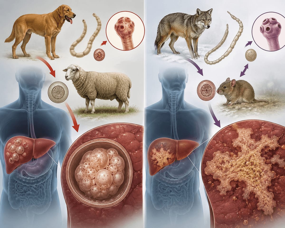
- AE is caused by Echinococcus multilocularis; CE by E. granulosus.
- On imaging, AE has no classic fluid-filled capsule — the lesion is infiltrative and multilocular.
- Oncology is often the first suspicion; differential diagnosis is essential.
- Without treatment, the AE prognosis is severe — do not delay scan review.
- A separate page covers cystic echinococcosis — if you have CE with a capsule.
How we work with patients from Kyrgyzstan
For patients in Kyrgyzstan, consistent step-by-step therapy is the core principle.
For patients from Kyrgyzstan, our priority is non-surgical care with continuous monitoring.
For cases from Kyrgyzstan, we focus on practical clarity: what to do first and how to measure progress.
Our cases from Kyrgyzstan show that early diagnostic validation lowers the risk of prolonged complications.
For patients in Kyrgyzstan, follow-up after the main course is included to stabilize outcomes.
How to reach Visus Medical: for patients from Kyrgyzstan
Из Бишкека до Ташкента — регулярные рейсы около 1,5 часов; перелёт выполняют несколько авиакомпаний, цены доступнее, чем из других столиц СНГ. Такси от аэропорта до Visus Medical — ещё около часа.
В Кыргызстане эхинококкоз встречается чаще среди жителей сельских районов с отарами — многие пациенты из Бишкека узнают о кисте на плановом УЗИ. Мы помогаем интерпретировать уже имеющиеся снимки без повторной диагностики.
Граждане Кыргызстана въезжают в Узбекистан без визы по паспорту — сухопутные переходы и авиа. Пограничный контроль стандартный, очереди обычно небольшие.
Alveococcosis: what patients from Kyrgyzstan should know
Shepherd camps and dogs in the mountains of Kyrgyzstan are a typical background in stories from patients from Kyrgyzstan.
Yellowing skin with a growing focus in a patient from from Kyrgyzstan is a reason for urgent scan review, not waiting for it to «pass».
MRI outside capitals of Kyrgyzstan is limited. Re-review of existing CT saves time and money.
We separate the alveococcosis route from common echinococcosis so the family understands timelines and goals.
Send CT, preferably MRI, from clinics in Kyrgyzstan — we give a preliminary answer on tactics.
Follow-up is tighter than for a usual cyst: that is built into the plan, without «later» surprises.
Hunting and life in high mountains in Kyrgyzstan are reasons to ask about alveococcosis directly with the doctor.
Patients from Kyrgyzstan are often referred to an oncologist for a liver focus without a clear capsule on the scan.
From Osh and Bishkek people bring a CT disc after hearing «surgery needed» — we first check whether tablets can be enough.
What is alveococcosis?: what patients from Kyrgyzstan should know
We advise patients from Kyrgyzstan not to interrupt the course — even when feeling better, checkpoints matter. Alveococcosis (alveolar echinococcosis, AE) is a severe parasitic disease of the liver. The parasite does not form a single bubble; it grows like a tumor, infiltrating tissue and sometimes producing distant foci. Evidence from Kyrgyzstan shows early therapy yields better outcomes. That is why CT often mistakes it for liver cancer.
Unlike cystic echinococcosis, “simply cutting out the cyst” is often impossible: the lesion has no clear borders. Conservative management and long-term follow-up are key topics when discussing treatment.
Symptoms and when to seek help
Early stages may have no complaints. Later, signs of liver and bile-duct involvement appear.
- Heaviness or pain in the right upper abdomen, loss of appetite, weakness.
- Obstructive jaundice if bile ducts are compressed or invaded.
- Weight loss, prolonged low-grade fever without a clear infection.
- On CT/MRI — an infiltrative lesion without a clear capsule; the report may say “suspected tumor.”
Diagnostics: why CT and MRI matter
For patients from Kyrgyzstan, we align the care plan with local logistics, test availability and follow-up timing. For Kyrgyzstan, avoiding diagnostic delays is especially important. For alveococcosis, ultrasound is only a starting point. The imaging “gold standard” is CT and/or MRI: they show infiltration borders and relations to vessels and ducts.
Serology helps but does not replace the imaging picture. Biopsy when AE is suspected is discussed cautiously — risk depends on location and clinic protocol.
- CT/MRI — extent of involvement and differentiation from tumor.
- Ultrasound — follow-up during treatment once a baseline CT exists.
- Antibody tests — supporting evidence, not the only argument.
AE treatment approaches in world practice
If you contact us from Kyrgyzstan, consultation format and treatment pacing are agreed in advance. International guidelines for AE more often discuss radical resection when technically feasible, and long-term antiparasitic therapy — especially for inoperable forms. Our protocol is a medication course with imaging control for patients suited to a non-surgical route.
- Surgery — when the lesion is resectable and the patient is ready for major intervention.
- Long-term medication therapy — the foundation when surgery is impossible or declined.
- Combined approaches — by multidisciplinary decision and imaging data.
- Watchful waiting without treatment in AE is generally unacceptable — unlike “inactive” CE4–CE5 cysts.
How alveococcosis is transmitted: what patients from Kyrgyzstan should know
Our approach for Kyrgyzstan focuses on a structured route without random protocol changes. The E. multilocularis cycle involves wild carnivores (foxes, wolves) and rodents. We advise patients from Kyrgyzstan to track their progress systematically. Humans become infected by swallowing eggs — via contaminated hands, berries, greens, or water in endemic zones.
Mountain and forest-steppe areas, hunting, and contact with hides and wild animals raise risk. This differs from the classic “shepherd — dog — sheep” epidemiology of cystic echinococcosis.
- Definitive hosts: foxes, wolves, sometimes dogs in endemic foci.
- Intermediate hosts: rodents; humans are accidental hosts.
- Prevention: hygiene outdoors, heat-treating forest foods, deworming dogs in endemic areas.
Prevention and what to do now — guidance for from Kyrgyzstan
For patients from Kyrgyzstan, early diagnostic verification and a properly structured therapy start are decisive. If your CT report already says “alveococcosis,” “alveolar echinococcosis,” or “infiltrative parasitic process” — send the scans. The earlier the review, the clearer the plan.
- Do not postpone CT/MRI review “for later.”
- Do not compare AE to an “ordinary cyst” — the tactic is different.
- If a tumor is suspected, clarify a parasitic nature before aggressive steps when timing allows.
- For families in endemic regions — hygiene measures and dog control.
Frequently asked questions (FAQ)
No, it is a parasitic disease. But on imaging it often mimics a tumor — so proper differential diagnosis is needed.
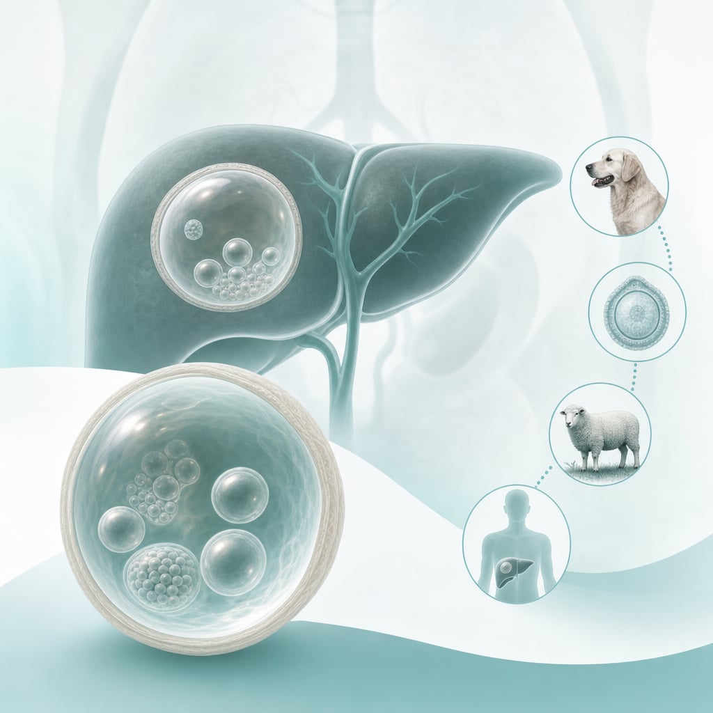
CE is a cyst with a capsule and fluid; AE grows infiltratively without clear borders. Treatment and prognosis differ; cystic echinococcosis is a separate service at our clinic.
In some cases — yes, with a medication protocol under imaging control. The decision comes only after CT/MRI review: send the files and we will say honestly.
Because AE looks aggressive and surgery often considers resection. A second opinion on your scans helps clarify whether a non-surgical option exists.
Usually longer than for a typical CE cyst. Duration is individual; the guide is dynamics on follow-up CT/MRI and ultrasound.
Yes. Without treatment, AE progresses and can lead to severe complications and death. Do not delay scan review.
Yes. Many come exactly when surgery is impossible or they are not ready for resection — we assess what can realistically be done conservatively.
Our course is usually outpatient. Hospitalization is discussed only if the clinical situation requires it.
Common questions from patients from Kyrgyzstan
Typically 2-6 weeks, depending on diagnosis, stage and treatment response.
Post-course monitoring: scheduled labs and remote consultations to sustain results.
Typically 2-6 weeks, depending on diagnosis, stage and treatment response.
Post-course monitoring: scheduled labs and remote consultations to sustain results.
Typically 2-6 weeks, depending on diagnosis, stage and treatment response.
Send CT. A parasitic process must be ruled in or out before major surgery. That is exactly what primary review is for.
Often yes. If quality is low — we say which study to add. We do not order extra procedures «just in case».
Different cause, different scan picture, usually tighter follow-up. We do not mix services so timeline expectations stay honest.