Treatment at Visus Medical: Proprietary non-surgical method
For patients from Ashgabat, we align the care plan with local logistics, test availability and follow-up timing. For Ashgabat, avoiding diagnostic delays is especially important. This is what matters: we do not tell everyone that "surgery is unnecessary." We say that for most cases of cystic and alveolar echinococcosis there is a documented, patented alternative — and we have been applying it since 1995.
The Visus Medical protocol is a medication course aimed at parasite death and gradual cyst regression under ultrasound and CT monitoring. Treatment is outpatient: no hospital stay, no general anesthesia, no months of recovery after an incision.
Consultations and treatment are provided by a physician certified by the Ministry of Health of the Republic of Uzbekistan — within permitted medical practice. We advise patients from Ashgabat to track their progress systematically. Cysts are most often found in the liver, but the protocol also applies to the lungs, kidneys, and other sites. Patients come from Russia, Kazakhstan, Kyrgyzstan, and Tajikistan — including after failed surgery and recurrence; many first send scans remotely.
- No general anesthesia or surgical incision.
- No removal of part of the liver or lung.
- No hospitalization — outpatient treatment.
- We work with post-surgical recurrence.
- Dynamic monitoring: ultrasound and CT at every stage.
- Any cyst location: liver, lungs, kidneys, and other organs.
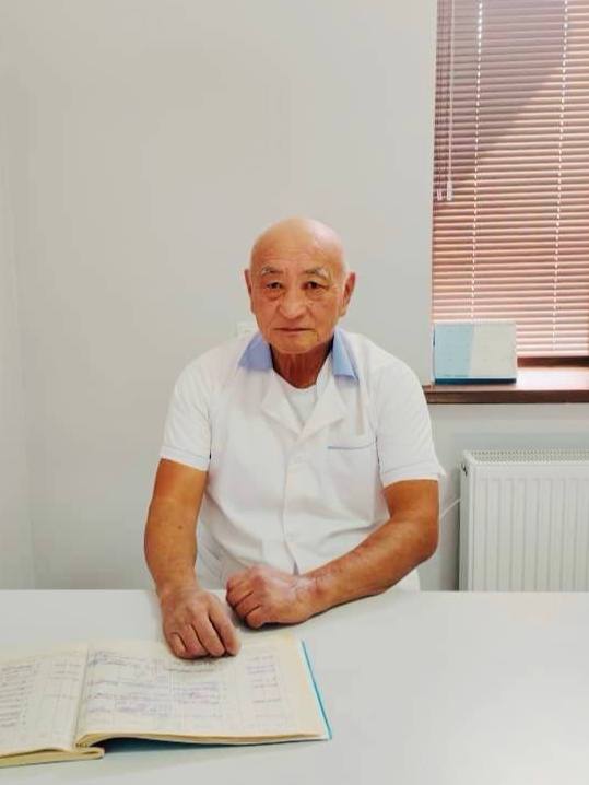
Elfréntiy Li
Chief physician — parasitologist, traditional medicine specialist
- 29 years treating echinococcosis and alveococcosis without surgery
- Higher School of Folk Medicine — licensed physician
- 600+ patients with documented results on follow-up imaging
- Proprietary non-surgical protocol in clinical use since 1995
“When you are told “surgery only,” a second opinion matters. We have treated echinococcosis without surgery for 29 years.”
Why do patients choose Visus Medical? — approach for Ashgabat
If you contact us from Ashgabat, consultation format and treatment pacing are agreed in advance. When a diagnosis feels like a sentence, it helps to know you are not obliged to accept the first option offered. Here is what sets us apart from the standard surgical route:
- MoH-certified physician in Uzbekistan — permitted medical practice, not unregulated folk care.
- 29+ years focused specifically on echinococcosis and alveolar echinococcosis — a specialty clinic, not one service among many.
- We handle complex cases: multiple cysts, alveolar echinococcosis, recurrence after surgery.
- Patients from 5 CIS countries — many come after being refused surgery or when it did not help.
- Transparent follow-up: imaging before, during, and after the course — you see the progress.
- Free initial consultation on your scans — send ultrasound or CT and we will assess your case before you decide.
How we work with patients from Turkmenistan
For cases from Ashgabat, we focus on practical clarity: what to do first and how to measure progress.
For patients in Ashgabat, consistent step-by-step therapy is the core principle.
For people in Ashgabat, our priority is non-surgical care with continuous monitoring.
For patients in Turkmenistan, follow-up after the main course is included to stabilize outcomes.
For patients from Turkmenistan, we usually begin with remote review of prior tests before planning the in-person phase.
Как добраться в Visus Medical: пациентам из Ashgabat
Для жителей Ashgabat оптимален двухдневный визит: день перелёта Ашхабад — Ташкент, день приёма и процедур в Visus Medical, обратный рейс на следующее утро или вечером того же дня при плотном графике.
Визовые требования для граждан Туркменистана при въезде в Узбекистан меняются — проверьте актуальные правила на момент поездки; клиника не оформляет визы, но подтверждает медицинскую цель визита письмом на русском языке.
Из Turkmenistan часть пациентов летят через Ашхабад с пересадкой: прямых рейсов в Ташкент из отдалённых городов мало, стыковка в столице Туркменистана добавляет полдня, но остаётся самым быстрым маршрутом.
Эхинококкоз: контекст для пациентов Turkmenistan
Гидатидные кисты печени нередко обнаруживают случайно при обследовании у пациентов from Ashgabat, которые годами не связывали дискомфорт в правом подреберье с контактом собак в сельской местности Turkmenistan.
Загрязнение почвы яйцами паразита у колодцев и овчарен в Turkmenistan — типичный сценарий заражения; мы объясняем пациентам from Ashgabat, как сочетать лечение с мерами профилактики для всей семьи.
Собаки без регулярной дегельминтизации в малых городах Turkmenistan остаются главным резервуаром паразита; дети, играющие во дворах рядом с дворами скота, попадают в группу повышенного риска.
Liver cyst on ultrasound or CT: could it be echinococcosis?: what Ashgabat residents should know
Our approach for Ashgabat and nearby areas focuses on a structured route without random protocol changes. We advise patients from Ashgabat to track their progress systematically. Most people do not arrive with a ready diagnosis of "echinococcosis" — the report says "hepatic cystic lesion," "parasitic cyst," or simply "liver cyst." That is normal: imaging finds the change first, then the cause is clarified.
A parasitic cyst in cystic echinococcosis usually appears as a round fluid-filled lesion with a capsule; sometimes daughter cysts are seen inside ("matryoshka" sign). In alveolar echinococcosis the picture differs: no clear capsule, the lesion looks infiltrative with multiple cavities — so oncology is often suspected first and surgery is offered quickly.
To avoid confusing it with a simple biliary cyst or benign tumor, you need combined diagnostics (ultrasound + CT or MRI + antibody testing) and an experienced specialist. Evidence from Turkmenistan shows early therapy yields better outcomes. We start with a free review of your scans — you can send a report from Almaty, Astana, Moscow, or any other city before traveling to Tashkent.
- If surgery was offered immediately — consider a second opinion on your scans before consenting.
- Treatment of hepatic echinococcosis at our clinic starts after WHO cyst staging (CE1–CE5), which defines the tactic.
- A still-small cyst — the earlier the course starts, the shorter and more predictable the follow-up ultrasound course.
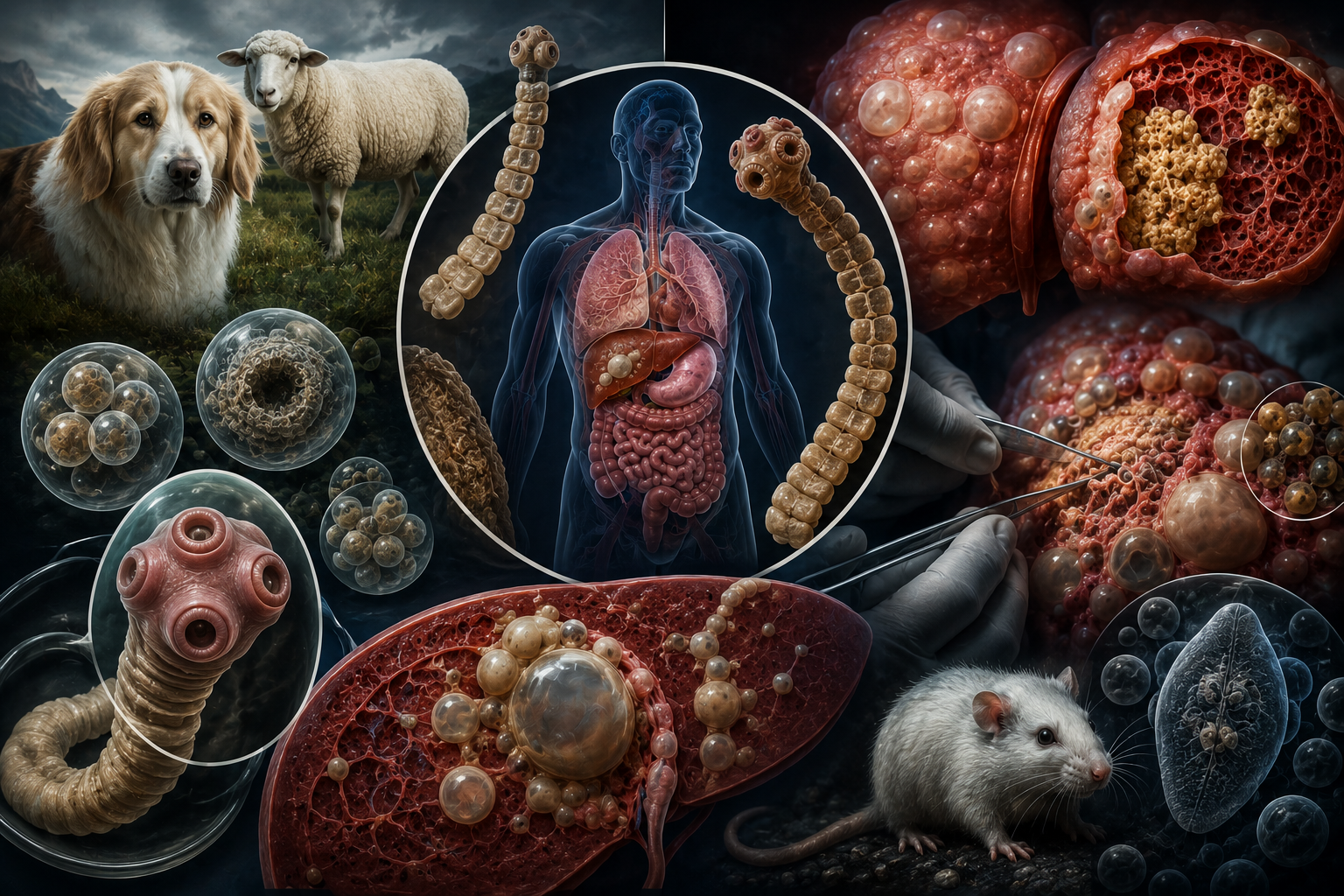
What is echinococcosis?
Echinococcosis is a serious parasitic disease: tapeworm larvae form cysts in the liver (in most cases), lungs, and other organs. Cysts can grow silently for years, then cause severe complications — including rupture with anaphylactic shock. The good news: with the right approach, the disease can be treated without surgery.
Two different threats: Cystic and alveolar echinococcosis
For residents of Ashgabat, we adapt the protocol to local realities — from logistics to repeat test access. Evidence from Turkmenistan shows early therapy yields better outcomes. It is extremely important to distinguish between the two main forms of the disease, as they follow different courses and require different treatment approaches:
Alveolar echinococcosis often mimics advanced malignancy and is considered one of the most dangerous helminthiases in humans.
- Cystic echinococcosis (CE), caused by Echinococcus granulosus. This is the most common form. The parasite forms one or more cysts (fluid-filled sacs) that grow slowly, like a "balloon" filled with fluid, displacing organ tissue.
- Alveolar echinococcosis (AE), caused by Echinococcus multilocularis. This form is less common but progresses much more aggressively. The parasite does not form a distinct cyst; instead it grows by infiltrating liver tissue, similar to a malignant tumor, and may produce "metastases" to other organs.
Symptoms: A silent enemy
If you live in Ashgabat, we can offer a hybrid format: remote stages + in-person checkpoints. The insidious nature of echinococcosis lies in its long asymptomatic period. A cyst can grow in the body for 5, 10, or even 15 years without causing any symptoms. The person feels completely healthy.
The first symptoms appear when the cyst reaches a significant size and begins to compress adjacent organs or ducts:
An acute complication is cyst rupture. This may occur spontaneously or after trauma. Cyst contents, which are highly allergenic, spill into the abdominal or thoracic cavity, which can cause severe anaphylactic shock (including cardiac arrest) and dissemination (spread) of the parasite throughout the body.
- When the liver is affected: Dull, aching pain or a feeling of heaviness in the right upper quadrant, nausea, loss of appetite. If the cyst compresses the bile ducts, mechanical jaundice may develop (yellowing of the skin and sclera).
- When the lungs are affected: Chest pain, shortness of breath, prolonged dry cough, sometimes hemoptysis.
- General symptoms: Allergic reactions (urticaria, pruritus), general weakness, fatigue, weight loss.
Diagnosis of echinococcosis and alveolar echinococcosis: ultrasound, CT, blood tests: what Ashgabat residents should know
We advise patients from Ashgabat not to interrupt the course — even when feeling better, checkpoints matter. For Ashgabat, avoiding diagnostic delays is especially important. Diagnosis is not a single test but a chain: imaging shows the cyst and its type, serology confirms contact with the parasite, and the physician links this to history and stage. At the first consultation we usually work with what you already have — ultrasound, CT, or MRI from your local clinic.
The "gold standard" combines imaging and laboratory tests. Diagnosing alveolar echinococcosis especially requires CT or MRI — without them, infiltrative disease is easily mistaken for cancer.
Biopsy (needle aspiration of the cyst) for diagnosis is generally not performed because of the high risk of rupture and dissemination.
- Ultrasound: The most accessible, safe, and informative method for primary diagnosis. It not only detects a cyst but also stages it according to the WHO international classification (WHO-IWGE), which divides cysts into active (CE1, CE2), transitional (CE3), inactive (CE4), and dead (CE5).
- CT and MRI (computed and magnetic resonance tomography): Used to refine the diagnosis, determine exact location, size, relationship to vessels and ducts, and when planning treatment. They are especially important in alveolar echinococcosis to assess the degree of tissue invasion.
- Serological tests (blood tests): Detection of specific antibodies to echinococcus (ELISA, indirect hemagglutination). A positive result confirms contact with the parasite. However, a negative result does not always rule out disease (for example, with an isolated cyst or at early stages).
Treatment approaches in international practice — guidance for from Turkmenistan
For referrals from Ashgabat, we emphasize transparency: every stage has a clear purpose and expected outcome. Treatment strategy depends on the type (CE or AE), size, location, and activity stage of the cyst. The following approaches are used in international practice:
- Observation ("Watch and Wait"): Used only for inactive, "dormant," or dead cysts (CE4, CE5) that cause no symptoms and carry no risk of complications.
- Drug therapy: Long-term antiparasitic drugs (for example, albendazole). Used as the main method for inoperable cysts, to prevent recurrence after surgery, or to prepare for surgery.
- PAIR (Puncture, Aspiration, Injection, Re-aspiration): A minimally invasive method in which, under ultrasound guidance, the cyst is punctured, fluid is aspirated, alcohol or another agent is injected into the cavity to kill the parasite, and then aspirated again. Not applicable to all cyst types.
- Surgery: The traditional method, especially for giant cysts, complications, or alveolar echinococcosis. This is complex surgery requiring removal of the cyst with its capsule, and sometimes resection (partial removal) of the organ.
Parasite life cycle: How does infection occur?
Echinococcus has a complex life cycle involving two hosts:
Humans are accidental intermediate hosts. We are infected not from sheep or cattle but in the same way they are—by swallowing parasite eggs. This happens:
In the human digestive tract, an egg releases a larva (oncosphere) that penetrates the intestinal wall, enters the bloodstream, and is carried—most often to the liver or lungs—where it develops into a cyst.
- Definitive hosts: Carnivores (dogs, wolves, foxes). Adult worms live in their intestines and produce thousands of eggs shed in feces into the environment.
- Intermediate hosts: Herbivores (sheep, cattle, goats) and rodents. They become infected by eating grass contaminated with eggs. Larvae (cysts) develop in their organs (liver, lungs).
- Through contact with infected dogs (eggs may be on their fur or tongue) followed by poor hygiene (unwashed hands).
- By eating unwashed vegetables, berries, and greens from gardens where infected animal feces may be present.
- By drinking water from contaminated sources.
- When skinning wild predators (for hunters).
Prevention: How to protect yourself and your family?
If you contact us from Ashgabat, consultation format and treatment pacing are agreed in advance. Knowing the routes of infection leads to simple but effective prevention rules:
If you already have a diagnosis — do not delay. An echinococcal cyst does not resolve on its own: the smaller it is, the simpler and shorter the course. Evidence from Turkmenistan shows early therapy yields better outcomes. Contact us — we will review your case.
- Hand hygiene: Wash hands thoroughly with soap after any contact with dogs (even pets), after working in the garden or yard, and always before eating.
- Deworming pets: Give dogs anthelmintic drugs prescribed by a veterinarian regularly (every 3–4 months).
- Food safety: Wash all greens, vegetables, fruits, and berries thoroughly under running water, preferably boiled water, especially those picked from the garden.
- Clean water: Do not drink water from open, unverified sources (streams, wells) that may be contaminated with feces.
- Caution: Limit contact, especially for children, with stray and unsupervised dogs.
Frequently asked questions (FAQ)
No. Humans are a dead-end accidental host. Infection occurs only by swallowing helminth eggs shed by definitive hosts (for example, dogs).
Not directly, but the risk exists and should be controlled: deworm your dog every 3–4 months as recommended by a vet, wash hands after contact, and do not allow the dog to lick your face.
Yes. A large share of our patients had cysts return after surgical treatment. That is one reason we focus on the medication method: it acts on the parasite systemically, not just by removing the "sac."
With cystic echinococcosis (CE), complete parasite destruction and cyst regression are achievable. Alveolar echinococcosis (AE) needs longer treatment and individual assessment. We never promise an outcome without reviewing your scans — send ultrasound or CT and the doctor will tell you what is realistic in your case.
It depends on type (cystic or alveolar), size, number, and activity of cysts. The Visus Medical protocol is tailored after your scans are reviewed. We track progress with ultrasound/CT so you see real change at each stage.
Cysts grow and compress organs — jaundice, portal hypertension, respiratory failure are possible. Cyst rupture can cause anaphylactic shock and spread of the parasite. Untreated AE almost always leads to a fatal outcome.
No. Liver cysts have many causes: simple, parasitic, biliary, and others. Echinococcosis is confirmed by ultrasound/CT features, antibody tests, and clinical context. If your report says "parasitic cyst" or "suspected echinococcosis," send your scans — we will help clarify before you commit to surgery.
In cystic echinococcosis (CE) you usually see a separate capsule and fluid content, sometimes daughter cysts. In alveolar echinococcosis (AE) there is no clear border; liver tissue appears infiltrated with multiple cavities — on CT this is often confused with cancer. Treatment tactics differ; AE usually needs longer follow-up.
Surgery is justified for acute complications (rupture, compression with sepsis), some giant cysts, and when medical options are exhausted. For many active CE cysts and some AE cases, international guidelines try conservative or minimally invasive treatment first. After reviewing your scans we will honestly say whether our non-surgical course fits you or a surgeon is needed.
Common questions from patients in Ashgabat
Yes. We usually start with remote case review, then schedule in-person visits and follow-up checkpoints.
Yes, a hybrid model: online consultations for adjustments + in-person checkpoints.
Yes. We usually start with remote case review, then schedule in-person visits and follow-up checkpoints.
Yes, a hybrid model: online consultations for adjustments + in-person checkpoints.
Yes. We usually start with remote case review, then schedule in-person visits and follow-up checkpoints.