Alveococcosis (alveolar echinococcosis, Echinococcus multilocularis) is a serious liver disease with infiltrative growth. Unlike a classic encapsulated echinococcal cyst, alveococcus spreads into liver tissue and can compress bile ducts, vessels, and nearby organs.
Jaundice is one of the most visible signs of complication. Skin and sclera turn yellow, urine darkens, stool may lighten. This is not always "hepatitis" — with alveococcosis the cause is often mechanical: the parasitic lesion blocks bile outflow.
In Central Asia classic echinococcosis is more common, but alveococcosis cannot be ruled out with ambiguous liver lesions. Understanding symptoms helps patients seek care in time.
Why jaundice develops
Bile is produced in the liver and flows through ducts into the duodenum. When an alveococcosis lesion compresses main bile ducts (common bile duct or intrahepatic ducts), bile cannot drain normally — bilirubin builds up in the blood.
Mechanical (obstructive) jaundice differs from viral hepatitis: it is often accompanied by itching, pale stool, and dark urine. Right upper abdominal pain may be constant or worsening.
Jaundice sometimes develops gradually over weeks or months. Sometimes it progresses faster if lesion growth accelerates or inflammation occurs in the compressed area.
- Bile duct compression by infiltrative growth.
- Bilirubin buildup — yellow skin and sclera.
- Dark urine, pale stool — typical obstruction signs.
- Skin itching — common with mechanical jaundice.
- Onset may be gradual or accelerated.
Bile duct obstruction and other complications
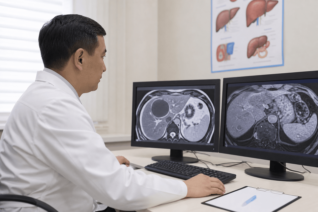
Beyond jaundice, alveococcosis can cause cholangitis (bile duct inflammation), liver abscess, diaphragm involvement, and metastasis-like spread to nearby structures. On CT and MRI it appears as a heterogeneous lesion with blurred borders.
Late stages may bring ascites (fluid in the abdomen), portal hypertension, and bleeding. These complications require hospitalization and specialized treatment — not home remedies.
Important: biopsy for alveococcosis diagnosis is generally avoided because of seeding risk. Diagnosis relies on imaging, serology, and clinical findings.
- Cholangitis — bile duct inflammation with obstruction.
- Liver abscess — purulent complication needing treatment.
- Nearby organ involvement with infiltrative growth.
- Ascites and portal hypertension — late complications.
- Biopsy — usually not indicated because of risk.
Why alveococcosis is more aggressive than cystic echinococcosis
A classic echinococcal cyst (E. granulosus) grows slowly, has a capsule, and often causes no complaints for years. Alveococcus (E. multilocularis) grows infiltratively — like a tumor, without a clear shell — and gradually invades more liver tissue.
That is why jaundice and obstruction are not rare with alveococcosis but a logical complication as the lesion grows. Prognosis with late diagnosis is much worse than with a simple cyst.
Early detection and combined treatment (surgery + long-term albendazole) improve outcomes. But treatment is more complex than for a simple cyst and requires experience with parasitic liver disease.
- Cystic echinococcosis — slow growth, capsule.
- Alveococcosis — infiltrative growth without capsule.
- Jaundice is a common alveococcosis complication.
- Late diagnosis — unfavorable prognosis.
- Treatment is harder: often surgery + long therapy.
Warning signs: when to seek urgent care
Seek medical care immediately for: worsening jaundice, severe right upper abdominal pain, fever with chills, vomiting, inability to eat, altered consciousness, bleeding.
If you already have a liver lesion and jaundice appears — do not delay workup. Urgent labs (bilirubin, alkaline phosphatase, GGT) and imaging (ultrasound, contrast CT or MRI) are needed.
When alveococcosis is suspected, specific serology (E. multilocularis) may be ordered. Routine E. granulosus ELISA can be negative — that does not rule out alveococcosis.
- Worsening jaundice — urgent reason to visit.
- Fever with right upper abdominal pain.
- Vomiting, weakness, loss of appetite with jaundice.
- Altered consciousness — emergency.
- Urgent labs and CT/MRI with new jaundice.
Urgent imaging: what is ordered
Ultrasound is the first step: it shows duct dilation, lesion size and structure. With jaundice, ultrasound is often done the same day.
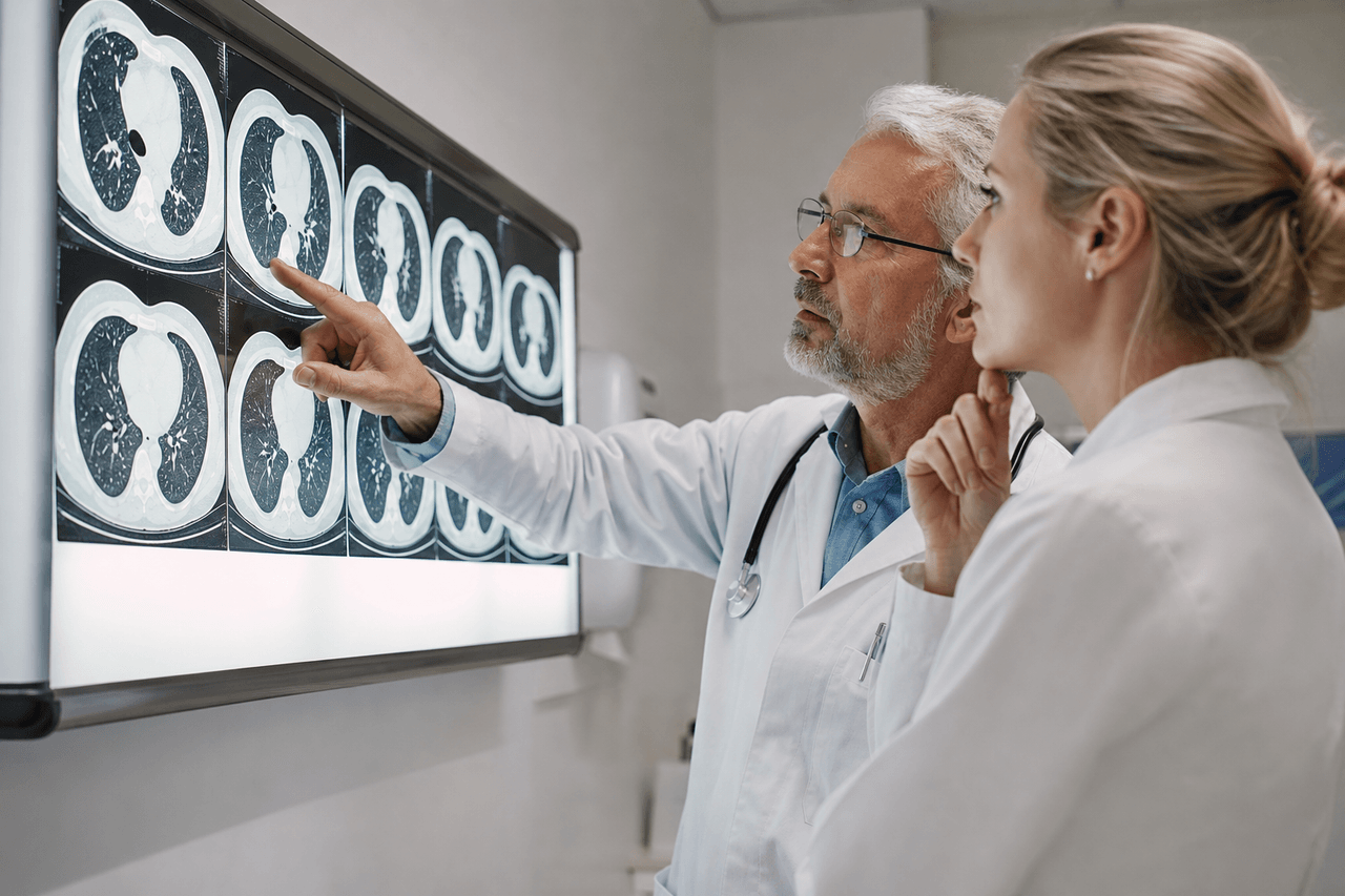
Contrast CT or MRI clarifies borders, relation to vessels and ducts, and differential diagnosis from liver cancer and cholangiocarcinoma. Alveococcosis often looks like a heterogeneous lesion with multiple small cavities.
PET-CT and other methods are used when indicated at specialized centers. The goal is not "another scan" but an accurate picture for treatment planning.
- Ultrasound — same day when jaundice is present.
- Contrast CT/MRI — for detailed assessment.
- Evaluation of bile ducts and vessels.
- Differential diagnosis from liver tumor.
- Serology — confirmation of parasitic origin.
Alveococcosis care at Visus Medical
At Visus Medical we specialize in liver echinococcosis and alveococcosis. With jaundice and suspected obstruction we promptly review images, order necessary imaging and serology, and explain what is happening and which steps are realistic for your case.
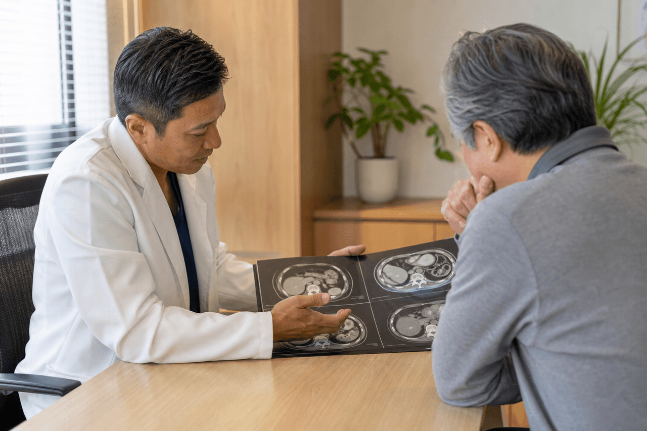
If you already have ultrasound or CT showing a "heterogeneous liver lesion" — bring images to the consultation. We distinguish classic cyst from alveococcosis, avoid unnecessary tests, and do not miss dangerous findings.
Alveococcosis is a serious disease, but with early care and correct management treatment options exist. We work with this daily in Central Asia and know what to look for first when jaundice appears.
Need an echinococcosis consult?
Message us on Telegram or WhatsApp — we’ll review your scans and suggest a plan.