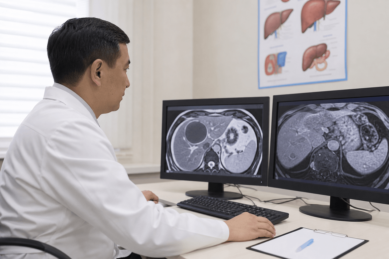
An echinococcal lung cyst may stay silent for years and first appear on chest X-ray, fluorography, or CT — at a checkup, before surgery, or during workup for cough.
Sometimes patients are treated for "bronchitis," "allergy," or "pneumonia" for months until the cyst grows and causes clear complaints. The lungs are the second most common site after the liver (about 15–20% of cases).
In Central Asia this is especially relevant: infection follows the dog–herbivore–human cycle and household factors — contact with dogs, unwashed greens, soil work. Lung echinococcosis often coexists with liver cysts — multiple organ involvement in 10–15% of patients.
Typical complaints
Symptoms depend on cyst size, location, and stage. A small peripheral cyst may be asymptomatic for years. As it grows, respiratory and general complaints appear.
Cysts near the lung root or central areas cause cough and shortness of breath earlier. Peripheral cysts stay "quiet" longer. If the cyst opens into a bronchus, contents may appear in sputum — sometimes described as bubbly or foul-smelling.
- Dry or productive cough, sometimes with blood streaks (hemoptysis).
- Chest pain or pressure, worse with deep breaths.
- Shortness of breath on exertion, air hunger.
- Fever when the cyst becomes infected (up to 38–39 °C).
- Allergic reactions when contents leak — hives, itching.
- General weakness, weight loss in long-standing disease.
- Right upper abdominal pain — if the liver is also involved.
How the cyst is found
Most often pulmonary echinococcosis is discovered incidentally on occupational chest X-ray. The film shows a round shadow with clear contours. Calcified walls may look like a dense ring.
X-ray gives initial information, but chest CT is the gold standard: exact size, location, number of cysts, relation to bronchi and pleura. Chest ultrasound is used when the cyst abuts the chest wall.
Along with lung imaging, abdominal ultrasound is always recommended to rule out liver and other organ involvement.
- Fluorography/X-ray — often first incidental finding.
- Chest CT — clarifies size, structure, number of cysts.
- Chest ultrasound — for peripherally located cysts.
- Abdominal ultrasound — mandatory to rule out liver cysts.
- Echinococcus antibody ELISA — confirms parasitic origin.
- Bronchoscopy — rarely, if bronchial rupture is suspected.
Features of pulmonary echinococcosis
Lung cysts grow more slowly than hepatic ones but carry their own risks. Rupture into the pleural cavity can cause hydatid hydrothorax — fluid between lung and chest wall. Bronchial rupture risks seeding and spread.
Cyst suppuration (abscess) presents with high fever, purulent sputum, and marked weakness. This requires hospitalization and active treatment.
With multiple lung and liver lesions, strategy is chosen comprehensively: which focus is more active, where complication risk is higher, what can be treated conservatively.
- Slow growth — but complication risk remains.
- Possible bronchial rupture with cough and sputum.
- Hydrothorax — pleural fluid after pleural rupture.
- Suppuration — fever, purulent sputum, weakness.
- Often combined with liver cysts — both organs must be assessed.
- Calcified cysts — inactive but need follow-up.
Treatment: surgery or medication
Pulmonary echinococcosis management depends on cyst size, location, and stage. Surgery was once the main method; today certain stages allow drug treatment with CT follow-up — similar to liver cysts.
Surgery is more often indicated for large cysts, rupture risk, infection, compression of nearby structures, or failure of conservative therapy. Modern video-assisted thoracoscopy allows less traumatic removal than in the past.
Drug treatment (albendazole and tailored regimens) is used for active cysts, multiple lesions, and as preparation for surgery. The decision is made after full workup.
Why delay is risky
Lung cyst rupture is dangerous because of seeding of the abdomen, pleura, and severe allergic reaction. The earlier the nature of the shadow on imaging is defined, the safer and calmer the strategy choice.
"Wait — it may resolve on its own" is unsafe for an active hydatid cyst. Cysts do not resolve spontaneously; without treatment they grow or calcify while keeping complication risk.
Any round shadow on chest X-ray in an endemic region needs clarification — rule out echinococcosis, tuberculosis, tumor. CT and serology help.
Why not to delay — and where to go
At Visus Medical we assess lung and liver foci together: one workup — a full picture of involvement. That matters because management of multiple cysts differs from a solitary lesion.
We choose treatment based on imaging, disease stage, and overall condition. If fluorography found a lesion — bring images to consultation. We explain what it may be, which tests are needed, and realistic treatment options.
Early diagnosis of pulmonary echinococcosis opens more options — including treatment without surgery at the right cyst stage.
Need an echinococcosis consult?
Message us on Telegram or WhatsApp — we’ll review your scans and suggest a plan.