Treatment at Visus Medical: proprietary non-surgical method — guidance for from Uzbekistan
For patients from Uzbekistan, early diagnostic verification and a properly structured therapy start are decisive. We do not tell everyone that “surgery is unnecessary.” For most cases of cystic echinococcosis there is a documented, patented alternative — and we have built experience since 1995.
The Visus Medical proprietary method is a medication course aimed at parasite death and gradual cyst regression under ultrasound and CT monitoring. For Uzbekistan, avoiding diagnostic delays is especially important. Treatment is outpatient: no hospital stay, no general anesthesia, no months of recovery after an incision.
Consultations are led by a physician certified by the Ministry of Health of Uzbekistan. Most often the cyst is in the liver — the method also applies to lungs, kidneys, and other organs. Patients come from Russia, Kazakhstan, Kyrgyzstan, and Tajikistan — including after recurrence; many first send scans remotely.
- No general anesthesia or surgical incision.
- No removal of part of the liver or lung.
- No hospitalization — outpatient treatment.
- We work with post-surgical recurrence.
- Dynamic monitoring: ultrasound and CT at every stage.
- Any cyst location: liver, lungs, kidneys, and other organs.
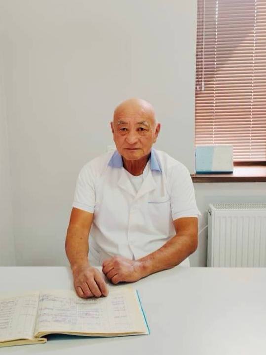
Elfréntiy Li
Chief physician — parasitologist, traditional medicine specialist
- 29 years treating cystic echinococcosis without surgery
- Higher School of Folk Medicine — licensed physician
- 600+ patients with documented results on follow-up imaging
- Proprietary non-surgical protocol in clinical use since 1995
“When you are told “surgery only,” a second opinion matters. We have treated echinococcosis without surgery for 29 years.”
Why patients choose Visus Medical
For residents of Uzbekistan, we adapt the protocol to local realities — from logistics to repeat test access. When a diagnosis feels like a sentence, it helps to know you are not obliged to accept the first option. For Uzbekistan, avoiding diagnostic delays is especially important. Here is what sets us apart from the standard surgical route:
- MoH-certified care in Uzbekistan — a physician authorized to practice.
- 29+ years focused on cystic echinococcosis — a specialty clinic, not one service among many.
- We handle complex cases: multiple cysts, giant CE, recurrence after surgery.
- Patients from 5 CIS countries — many come after surgery was refused or failed.
- Transparent follow-up: imaging before, during, and after the course.
- Free initial consultation on your scans — send ultrasound or CT before the visit.
How we work with patients from Uzbekistan
For patients from Uzbekistan, we build a clear plan with milestones, timelines and checkpoints.
Each patient from Uzbekistan receives a personalized route: consultation, diagnostics, treatment and follow-up.
Our team supports patients from Uzbekistan through each phase to keep treatment consistent.
For referrals from Uzbekistan, we evaluate comorbid factors and adapt treatment pace accordingly.
Our cases from Uzbekistan show that early diagnostic validation lowers the risk of prolonged complications.
How to reach Visus Medical: for patients from Uzbekistan
На авто из Tashkent до клиники Visus Medical в Нурафшоне — около часа по трассе через Ташкент и Чирчик; ориентируйтесь на выезд утром, чтобы избежать вечерних пробок на подъезде к столице.
Такси из Tashkent до Нурафшона обойдётся сопоставимо с поездкой до центра Ташкента; многие пациенты from Tashkent выбирают этот вариант за предсказуемость времени и комфорт после процедур.
Если вы живёте in Tashkent, можно совместить поездку в клинику с делами в Ташкенте: приём в Visus Medical займёт полдня, остальное время — на анализы или покупки в столице.
Если вы приезжаете from Tashkent с родственником, парковка у клиники Visus Medical бесплатная; заранее сообщите администратору время прибытия — мы подготовим кабинет и документы к вашему приезду.
Echinococcosis: what patients from Uzbekistan should know
In Uzbekistan sheep farming and yards with dogs are a common background for liver cysts. We ask patients from Uzbekistan about contact with livestock.
Tests and ultrasound can be done at local labs in Uzbekistan; we match results with what the scans show.
For residents of Uzbekistan a short route works well: consultation, starting the course, and follow-up ultrasounds without long logistics.
The first step is simple: photos or scan files in Telegram or WhatsApp. The doctor will say whether the method fits and what you need before the visit.
CT in regional centers of Uzbekistan is not available everywhere. We accept scans you already have — without unnecessary repeats when quality is good.
The course is outpatient: patients from Uzbekistan combine visits with work and home monitoring when possible.
Cyst recurrence after removal in clinics in Uzbekistan is a common reason to book a scan review with us.
At district clinics ultrasound often describes a cyst as a «liver lesion» without details. We clarify the picture from your scans.
At markets in Uzbekistan greens from the street and contact with animal fur are typical details in a patient story from from Uzbekistan.
Liver cyst on ultrasound or CT: how to recognize echinococcosis
Most people arrive without a ready diagnosis — the report says “hepatic cystic lesion,” “parasitic cyst,” or simply “liver cyst.”
In cystic echinococcosis, imaging usually shows a round fluid-filled lesion with a capsule; sometimes daughter cysts are inside (“matryoshka”). We advise patients from Uzbekistan to track their progress systematically. Stage is assessed by the WHO classification (CE1–CE5) — that guides the tactic.
To avoid confusing it with a simple cyst or another lesion, you need ultrasound + CT/MRI + serology and an experienced eye. You can send a report from Almaty, Astana, Moscow, or any city — before traveling to Tashkent.
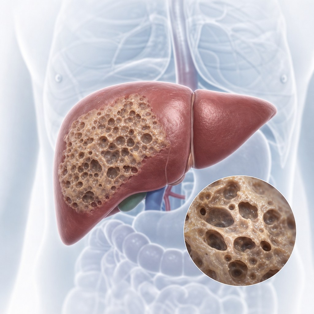
If imaging shows no clear capsule and the process looks infiltrative — it may be alveococcosis: we have a separate alveococcosis treatment service.
- If surgery was offered immediately — get a second opinion on your scans before consenting.
- Treatment starts after WHO cyst staging (CE1–CE5).
- The smaller the cyst, the shorter and more predictable the dynamics on follow-up ultrasound.
What is cystic echinococcosis?
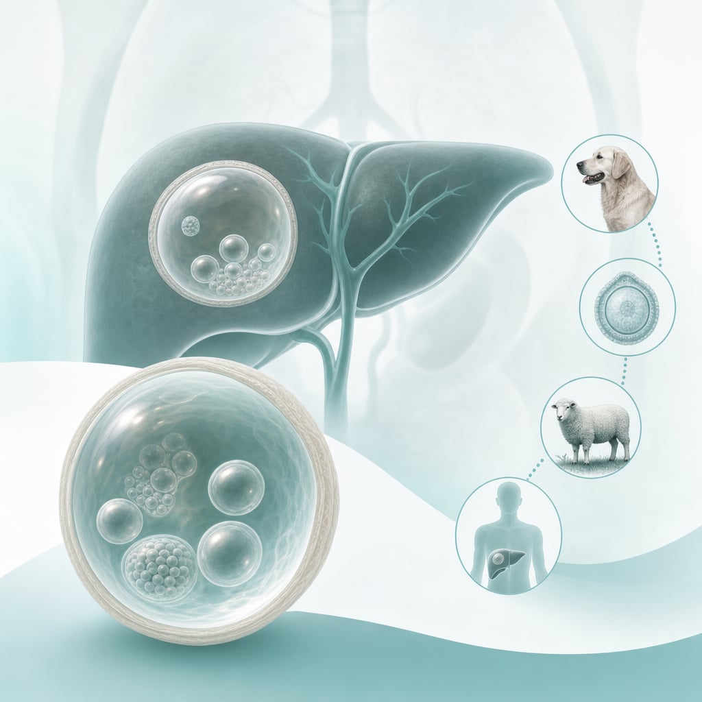
We advise patients from Uzbekistan not to interrupt the course — even when feeling better, checkpoints matter. Cystic echinococcosis (CE) is caused by Echinococcus granulosus. Larvae form one or several cysts in the liver (most often), lungs, and other organs. Cysts can grow for years without symptoms, then cause severe complications — including rupture with anaphylactic shock. We advise patients from Uzbekistan to track their progress systematically. With the right approach, the disease can be treated without surgery.
Symptoms: a silent enemy — guidance for from Uzbekistan
For referrals from Uzbekistan, we emphasize transparency: every stage has a clear purpose and expected outcome. The insidious nature of echinococcosis is a long asymptomatic period. A cyst may grow for 5–15 years without signs.
First symptoms appear when the cyst reaches significant size and compresses neighboring organs or ducts.
An acute complication is cyst rupture (spontaneous or traumatic): contents spill into the cavity, with possible anaphylactic shock and dissemination.
- Liver: dull pain or heaviness in the right upper abdomen, nausea, loss of appetite; bile-duct compression may cause obstructive jaundice.
- Lungs: chest pain, shortness of breath, dry cough, sometimes hemoptysis.
- General: urticaria, itching, weakness, fatigue, weight loss.
Diagnosing echinococcosis: ultrasound, CT, lab tests for patients from Uzbekistan
For patients from Uzbekistan, we align the care plan with local logistics, test availability and follow-up timing. Diagnosis is a chain: imaging shows the cyst and stage, serology confirms contact with the parasite, and the physician links this to the history. Evidence from Uzbekistan shows early therapy yields better outcomes. At the first consultation we work with what you already have — ultrasound, CT, or MRI.
Cyst biopsy for diagnosis is generally not performed because of rupture and seeding risk.
- Ultrasound — accessible primary diagnosis and WHO staging (CE1–CE5).
- CT and MRI — location, size, and relations to vessels and ducts.
- Serology (ELISA, IHA) — antibodies to echinococcus; a negative result does not always rule out disease.
Treatment approaches in world practice
If you contact us from Uzbekistan, consultation format and treatment pacing are agreed in advance. Tactic depends on size, location, and cyst activity stage (CE1–CE5):
- Watch and Wait: only for inactive CE4–CE5 without symptoms or complication risk.
- Medication therapy: long-term antiparasitic regimens — the basis of our approach at a suitable stage.
- PAIR: ultrasound-guided puncture with aspiration and sclerosant — not for all cyst types.
- Surgery: for giant cysts, acute complications, or when conservative tactics are exhausted.
Parasite life cycle: how infection happens
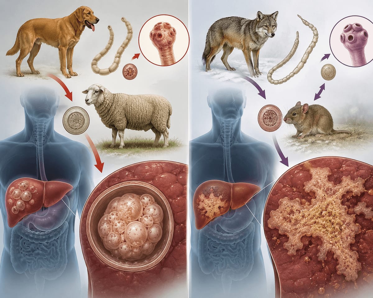
Echinococcus granulosus has a two-host cycle. For Uzbekistan, avoiding diagnostic delays is especially important. Humans are accidental intermediate hosts: infection by swallowing eggs (not directly from sheep).
In the intestine a larva emerges from the egg, enters the bloodstream, and most often settles in the liver or lungs, where a cyst develops.
- Definitive hosts: dogs and other carnivores — adult worm in the intestine, eggs in feces.
- Intermediate hosts: sheep, cattle, goats — cysts in organs.
- Human risk: contact with dog fur/tongue, unwashed vegetables and greens, water from contaminated sources.
Prevention
For patients from Uzbekistan, early diagnostic verification and a properly structured therapy start are decisive. If the diagnosis is already made — do not delay. A cyst does not resolve on its own: the smaller the size, the shorter the course. Write to us — we will review your case.
- Wash hands after contact with dogs and gardening.
- Deworm dogs every 3–4 months as advised by a veterinarian.
- Wash greens, vegetables, and berries thoroughly.
- Do not drink water from unchecked open sources.
- Limit children’s contact with stray dogs.
Frequently asked questions (FAQ) for patients from Uzbekistan
No. Humans are a “dead-end host.” Infection occurs only via eggs shed by definitive hosts (dogs and others).
There is risk if the dog is not dewormed. Regular treatment every 3–4 months and hand hygiene after contact reduce risk.
Yes. A large share of patients have cyst recurrence after surgery. The medication method acts systemically, not only by “removing the sac.”
In CE, full parasite destruction and cyst regression are achievable. We do not promise a result without reviewing scans — send ultrasound or CT.
Duration depends on WHO stage, size, and number of cysts. We assess dynamics with ultrasound/CT at every stage.
Cyst growth, organ compression, obstructive jaundice, respiratory failure. The most severe risk is rupture with anaphylaxis and seeding.
No. Cysts vary. Diagnosis combines ultrasound/CT, serology, and clinical picture. Send scans — we help clarify before a surgery decision.
Surgery is justified in acute complications, some giant cysts, and when medication tactics are exhausted. After reviewing scans we say honestly whether a non-surgical course fits.
Cystic echinococcosis is a separate cyst with a capsule. Alveococcosis grows infiltratively, like a tumor. These are different services: more on the alveococcosis treatment page.
Common questions from patients from Uzbekistan
Post-course monitoring: scheduled labs and remote consultations to sustain results.
Typically 2-6 weeks, depending on diagnosis, stage and treatment response.
Post-course monitoring: scheduled labs and remote consultations to sustain results.
Typically 2-6 weeks, depending on diagnosis, stage and treatment response.
Post-course monitoring: scheduled labs and remote consultations to sustain results.
Not always. Send ultrasound or CT in a messenger — we will review cyst stage and say whether data are enough for a plan or an in-person visit is needed. Many patients from Uzbekistan start this way.
Yes. Some follow-up ultrasounds are done at local clinics and files are sent to us. In-person visits follow the course schedule, without extra trips.
If the cyst fits tablet treatment — the course is done at home, without anesthesia and cuts. Surgery remains an option with complications and very large cysts — after scan review we say plainly what fits you.