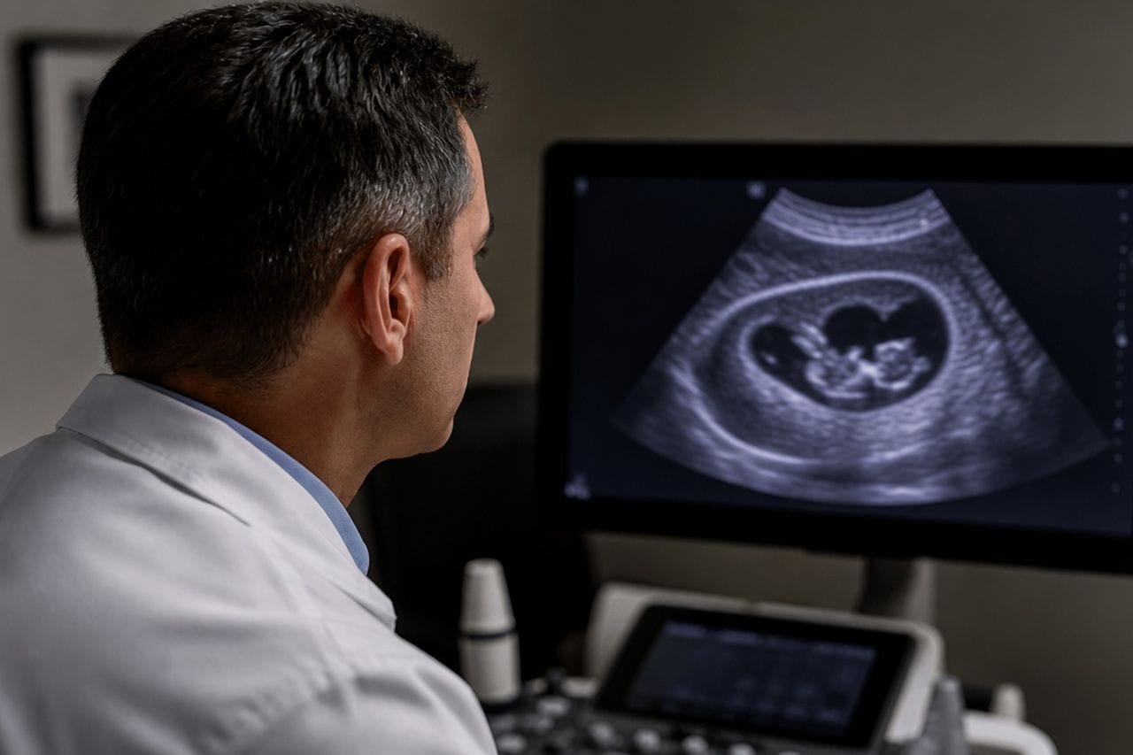
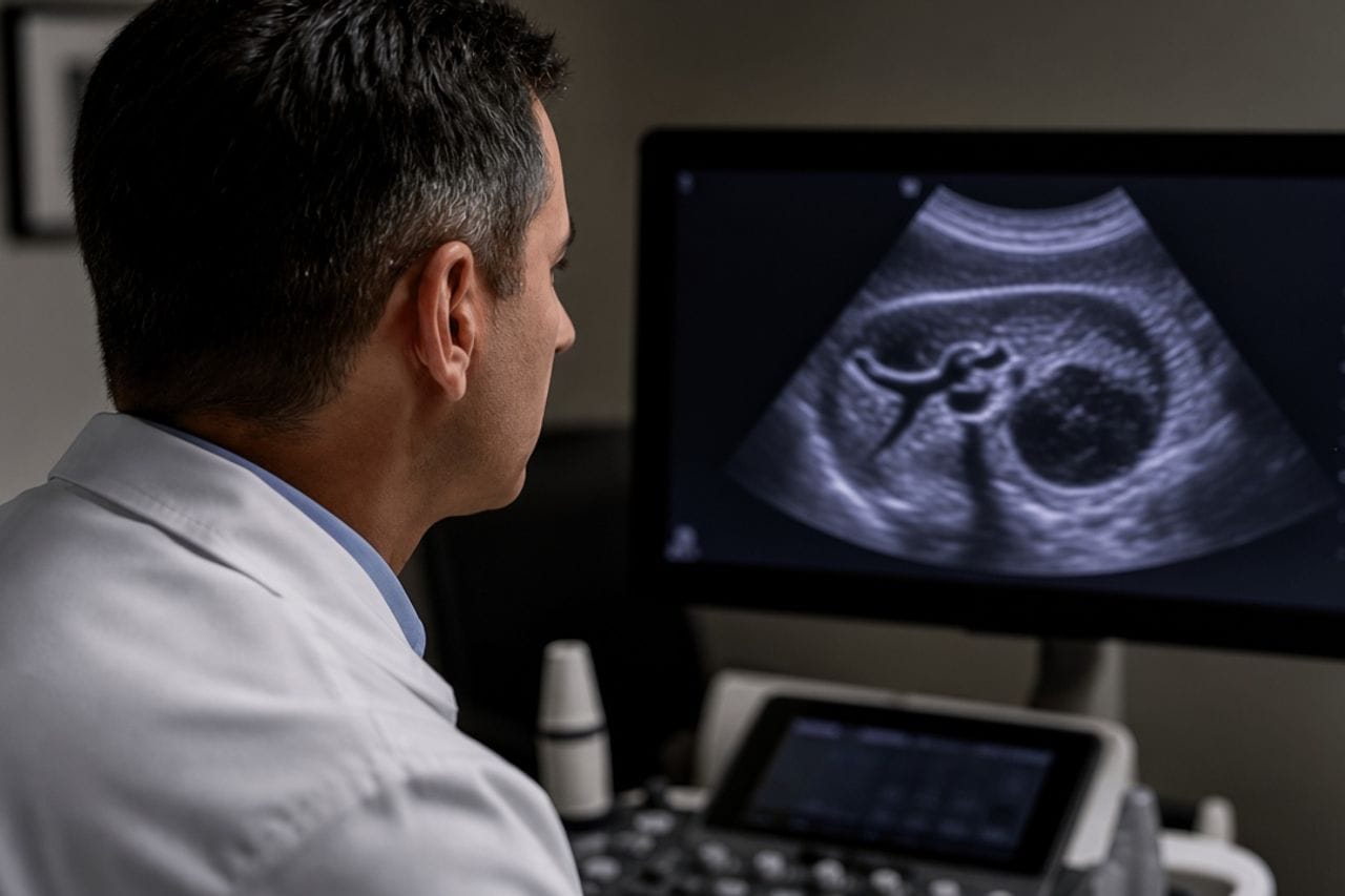
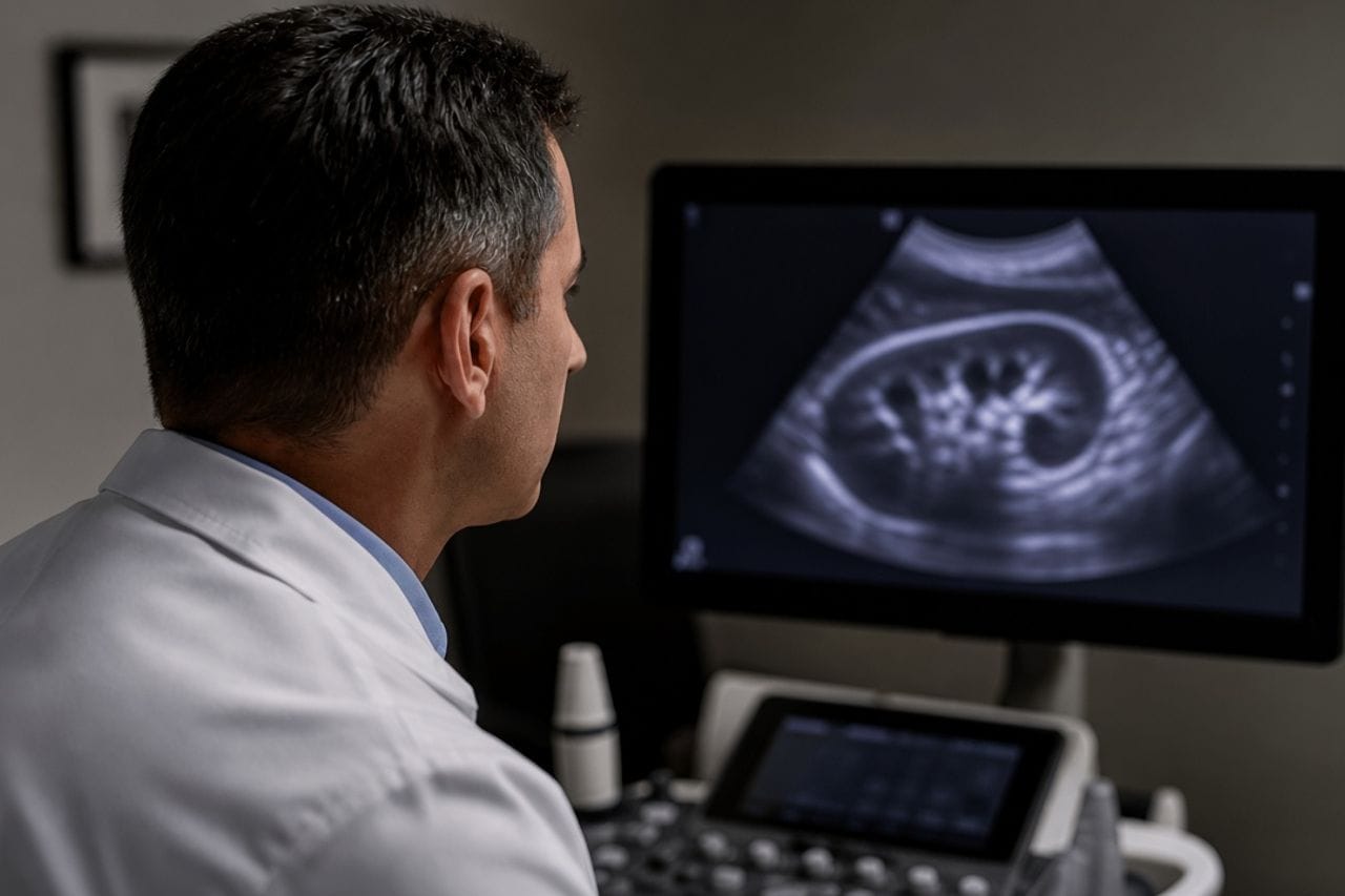
Alveococcosis starts in the liver. From there the process can give a distant lesion. The brain is one of those addresses, next to the lungs and less often the bones. On MRI this is a node or infiltrate that is easy to label as a tumor. A round cyst with a thin capsule, as in cystic echinococcus, is usually absent.
A person may treat the "liver" for a year and only then have a seizure. Or the reverse: a neurosurgeon finds a lesion in the head, and the liver has not yet been looked at. Both routes lead to one rule — image both the head and the liver, and do not choose only one.
A round cyst in the brain is a different disease: brain echinococcus. A lesion in the chest with the same alveococcosis: lungs. The letter M in the stage is exactly about these distant points: PNM.
Which complaints lead to MRI
Headache that builds over weeks, morning vomiting, seizures, weakness in an arm or leg, a change in speech, vision, or behavior. Some symptoms are blamed on blood pressure and cervical osteochondrosis until an attack happens.
Complaints from the liver can be modest at the same time: heaviness, weight loss, or none at all, although the liver lesion is already large. The brain sometimes announces itself louder than the liver. Symptoms of hepatic alveococcosis: what to watch for.
A sudden rash and abdominal pain do not belong to this article. That is how cyst rupture behaves, not an infiltrate in the brain: rupture.
- Seizures and rising headache with a liver lesion — MRI, not "vessels."
- Ultrasound does not show the brain. MRI is needed.
- The liver is imaged even if you came only because of the head.
- One normal neurological exam does not cancel a lesion on MRI if an attack has already happened.
- In children the same symptoms are not written off as school: alveococcosis in children.
How it looks on scans
The lesion is more often irregular in shape, may take up contrast in different ways, sometimes with edema around it. A tumor, tuberculoma, and abscess enter the same list, and the radiologist honestly writes "to differentiate." The word "alveococcosis" appears when there is a typical liver nearby or an already known diagnosis.
Head CT will see the lesion more coarsely. MRI is needed for a plan. PET and other methods are sometimes added by oncologists if the tumor version is still alive. They do not replace reading of the liver CT.
A brain biopsy "to find out faster" with already typical liver alveococcosis is not a starting step. A puncture carries risk, and the diagnosis is often assembled from the liver and the head together.
How management differs from a liver cyst
Pressure inside the skull does not wait for the scheme "small lesion, we will look in six months." A neurologist and neurosurgeon decide whether pressure must be relieved, the lesion removed, or its size watched on repeat MRI. This is not a copy of the decision for a hepatic CE1 cyst.
The liver is not abandoned at the same time. A distant lesion means the process is systemic. A drug course, if it is indicated, is prescribed by a doctor with both addresses in mind. There are no doses or duration in the article.
Confusing this lesion with an echinococcal brain cyst is dangerous in both directions. They try to take a cyst out entirely without opening it. An alveococcosis infiltrate does not "shell out" that way. The neurosurgeon's tactics depend on what is on the scan.
What else to check besides the head
CT or MRI of the liver with vessels is mandatory, even if ultrasound a year ago was "almost normal." The chest — because in alveococcosis the lungs are involved no less often than the head, and sometimes earlier.
Liver tests and antibodies complete the packet, but a negative ELISA with two typical lesions does not collapse the diagnosis. Serology: blood test.
If the liver has already been operated on, the old discharge is needed: whether the resection margin was taken for a "new" process, and whether the brain was under-examined before surgery. Follow-up after resection: after surgery.
When it is urgent, and when it is planned
A first seizure, rising weakness in a limb, vomiting, confusion, sudden severe headache — the same day. This is not a neurology appointment in three weeks.
An already known small lesion without new complaints is managed on an MRI schedule the doctor set. Skipping follow-up because "the head does not hurt" is a typical way to learn about growth too late.
Planned, but without self-reassurance: any new neurology in a person with liver alveococcosis, even if last summer the MRI was clean. A distant lesion can appear later.
- Do not stop anticonvulsants yourself if they have already been prescribed.
- Do not agree to surgery for a "brain tumor" until the liver has been reread.
- Bring discs of the head and liver to one table, not in different months.
- Ask whether your stage has the letter M1 or the head has not yet been looked at.
- A repeat MRI is compared with the previous one, not described from scratch.
Review at Visus Medical
Send head MRI and liver CT. We say whether this looks like alveococcosis and whom to see first: a neurosurgeon because of pressure, or first liver tactics if the head is stable.
If two clinics wrote different things — tumor and parasite — that is exactly the scan packet that is needed, before treatment of the wrong disease starts.
Alveococcosis. A report without an MRI disc is weak for this question: edema and the shape of the lesion sit in the series, not in two lines.
Need an echinococcosis consult?
Message us on Telegram or WhatsApp — we’ll review your scans and suggest a plan.