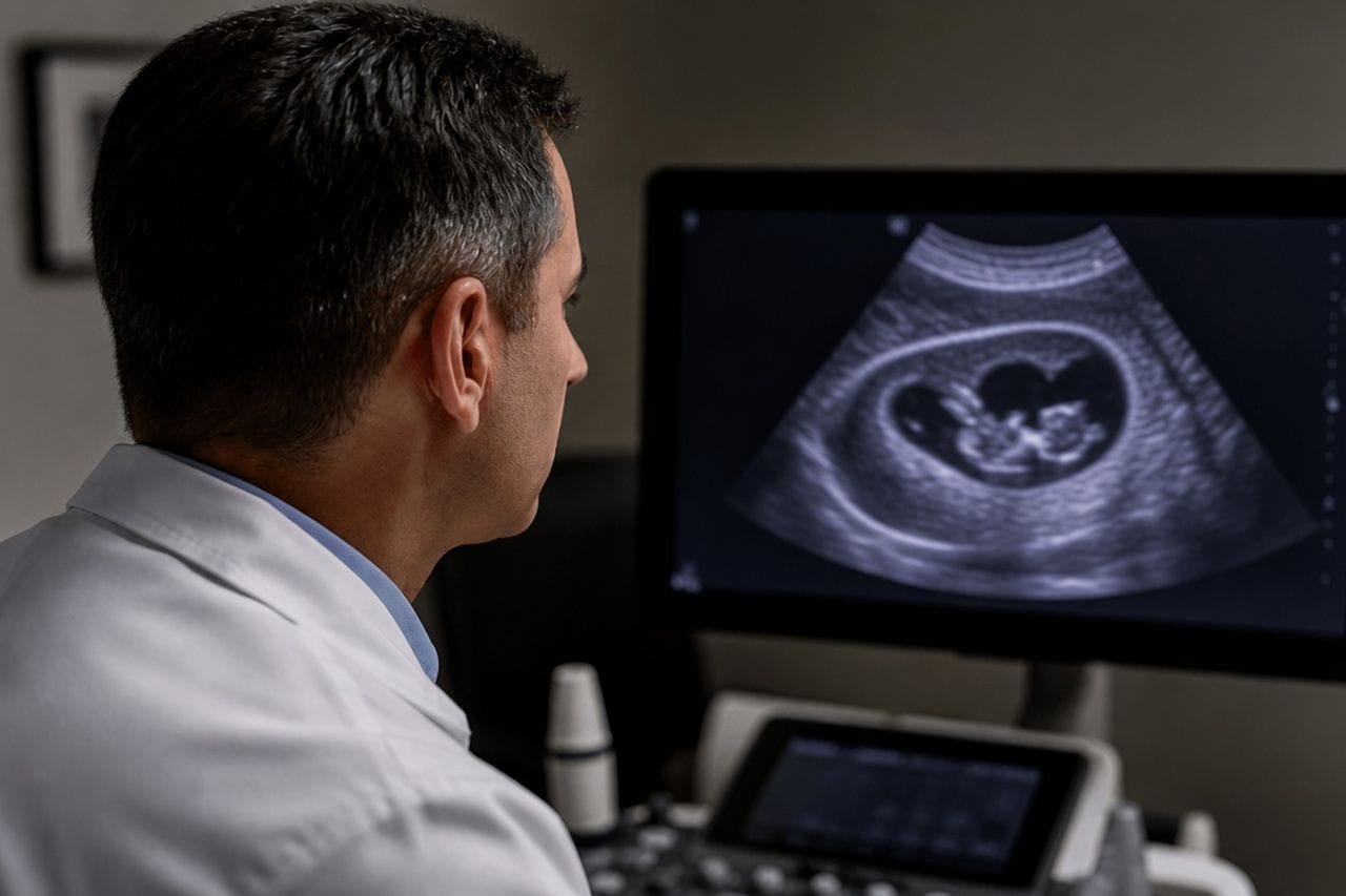
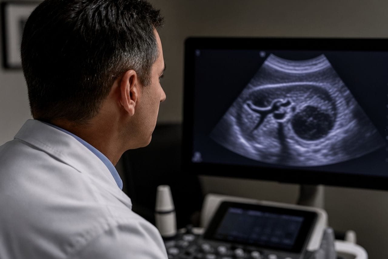
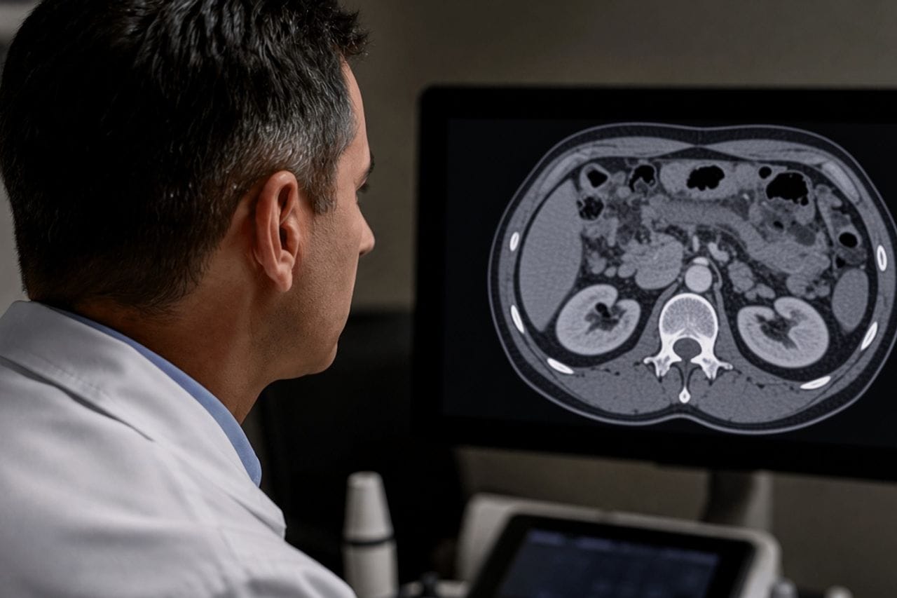
An intact echinococcal cyst is often silent. When infection enters the cavity, fever, chills, and pain in the side appear. On the scan the wall thickens, the contents become thick, and septa appear. The wording "liver abscess" at that moment sounds convincing and is sometimes incomplete.
An ordinary abscess and a suppurating echinococcus are treated differently. A blind puncture and drain, appropriate for a banal pus collection, can spread the contents around with a parasitic cyst. So a label on ultrasound is a reason to clarify, not to puncture at once.
Suppuration happens on its own and together with rupture into the bile ducts. Then jaundice is added to the fever. That is already two complications at once: rupture into the ducts.
Which complaints are typical
Fever, often with chills, pain or a bursting feeling on the right, weakness, sometimes nausea. The pain may be tolerable by day and keep you from sleeping at night. Some people take antipyretics for several days and blame it all on a virus.
If the cyst was already found earlier, comparison with an old ultrasound matters more than one fresh frame. A quiet anechoic cavity that suddenly became heterogeneous against a background of fever is a strong argument for suppuration, not that "the cyst just grew."
Sudden pain in minutes, a rash, and a drop in blood pressure are closer to rupture, even if fever is also present. Then the ambulance comes first: cyst rupture.
- Fever with pain in the right side — look today, not after the holidays.
- Bring old ultrasounds: the dynamics of the contents decide more than size.
- Do not agree to a puncture until a parasitic cyst has been excluded.
- Jaundice on a background of fever is a reason to look at the ducts as well, not only the "abscess."
- A blood test for antibodies does not replace CT at the height of fever.
How they distinguish it from an ordinary abscess
An abscess is more often linked to cholangitis, appendicitis, trauma, or another clear infection. Echinococcus is linked to an already known cyst or to life in an endemic region, even if the person was not thinking about that. On CT a parasitic cavity often has its own capsule, daughter vesicles, or calcification in the wall, which a fresh abscess does not have.
Serology helps when it is positive, and almost does not help when it is negative: in suppuration and with an intact cyst ELISA can be falsely negative. You cannot rely only on the antibody form. More detail: blood test.
A tumor with breakdown can also look "dirty" on ultrasound. If the contours are irregular and there is no confident capsule, the differential list includes both alveococcosis and a neoplasm. Then CT is needed before any drain: echinococcosis or cancer.
Why puncture is not the first step
A needle in a parasitic cyst without preparation and without understanding the stage can carry scolices into a duct or the abdominal cavity. For some stages there are their own minimally invasive methods; they are done in settings ready for such contents. This is not a procedure "in passing" in an ultrasound room.
First the picture: ultrasound plus CT, assessment of the ducts, signs of rupture, the number of cysts. Then the decision to drain, operate, or combine with a course. An article does not replace this choice and does not describe doses.
WHO stage is harder to read in suppuration: pus masks daughter vesicles. They still try to assess it, because it decides what happens after the fever falls: CE1–CE5.
How tactics differ from a quiet cyst
An inactive calcified cyst without complaints is often observed. Suppuration cancels that logic for the time of the acute infection. "Let us wait six months" with fever and pain in the side does not fit, even if a year ago the cyst was called CE5.
After the fever settles the cyst does not disappear by itself. A cavity remains that must be reviewed: is it active, is it connected to a duct, is there a second cyst in the lung. The liver and chest in echinococcus are looked at together more often than the patient expects.
Multiple cysts do not all suppurate at once. A map of lesions is needed so one "abscess" is not treated while the rest are missed: several cysts.
What to keep for the doctor
All ultrasounds from past years, the latest blood test with leukocytes and liver chemistry, discharge summaries if IV drips were already given. Write down how many days there was fever and whether there was jaundice.
If a drain was already placed in another clinic, you need the protocol: what was obtained from the cavity and whether material was sent for testing. That decides whether it was a parasite or ordinary pus.
Taking antiparasitic tablets on your own against high fever and without scans only confuses the liver picture. The drug, if it is needed, is added when it is clear what is happening with the cavity and the ducts.
- Do not heat the side and do not apply "resolving" compresses.
- An antipyretic does not cancel an examination; it only masks the temperature.
- A repeat rise in fever after discharge is a reason to return, not to endure.
- Ask whether a connection with the bile ducts was excluded.
- A follow-up scan after the fever falls is scheduled, not "when there is time."
Review at Visus Medical
Send ultrasound or CT and say how many days there was fever and whether a puncture was done. We separate echinococcus suppuration from an ordinary abscess and say whether a surgeon is needed at once or first further investigation.
If the acute period is already over, we look at what is left of the cyst and whether you can go further without repeated openings.
Echinococcosis. Bring old discs: one "abscess" on a fresh ultrasound without an archive is easy to read wrongly.
Need an echinococcosis consult?
Message us on Telegram or WhatsApp — we’ll review your scans and suggest a plan.