Cystic echinococcus has a cavity and a wall. If the wall communicates with a bile duct, fluid and scraps of membrane enter the pathway. The duct clogs, bile cannot exit, and the person gets an attack that is easy to label as stone disease.
A connection between the cyst and a duct can be quiet: mild bitterness, episodes of dark urine, "liver" pain without bright jaundice. It can also be acute: colic, jaundice, and chills in the same day. Both versions are not a reason to take a choleretic and wait a month for a planned ultrasound.
Alveococcosis also causes jaundice, but the mechanism is different: the infiltrate compresses the ducts from outside; there is no bubble with a wall. Mixing up these two texts is dangerous, because the urgent steps do not match. About compression: alveococcosis and jaundice.
How it feels
The classic picture is sudden pain in the right upper abdomen, pale stool, dark urine, itching of the skin, then yellowing of the sclera. Fever is added when inflammation of the ducts joins in. Nausea is blamed on "fatty food yesterday."
If a liver cyst was already in an ultrasound report, that detail must be told to the doctor in the first minute. Otherwise they look for a stone in the gallbladder and may not look at the hepatic ducts higher up.
A rash, swelling, and a drop in blood pressure the same night mean it is not only the duct: they also think of rupture into the abdominal cavity. That is already an ambulance without a pause: cyst rupture.
- Jaundice with pain does not wait for a planned ultrasound next week.
- Name the known liver cyst, even if it was found long ago and "it is quiet."
- Fever plus jaundice is inflammation of the ducts, not a cold.
- Choleretics and warming do not sort out what is lying in the duct.
- Bring old scans with you: without them it is not clear whether a connection with a duct was there earlier.
What investigations show
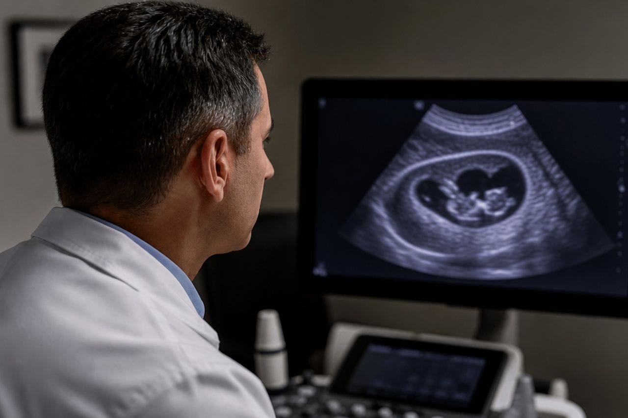
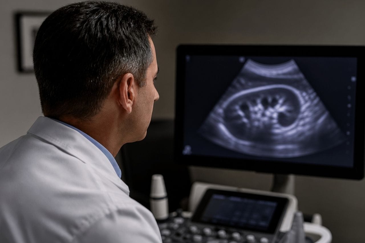
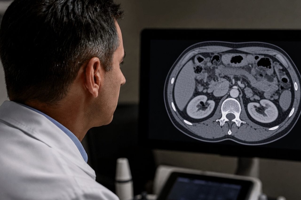
Ultrasound can show dilated ducts and the cyst itself, but scraps of membrane inside a duct often are not proven by it. CT or MRI of the liver clarifies whether there is a communication, how dilated the ducts are, and whether there are other cysts.
Sometimes the ducts are examined from inside to remove what is plugging them and restore bile flow. This is not a "test out of curiosity" but a way to relieve the block. The decision is made by the doctor who sees the scans, not by an article.
An antibody test for echinococcus is secondary on such a night. A negative ELISA does not cancel a cyst that is already visible. How to read serology in a calm situation: blood test.
How this is not alveococcosis
In alveococcosis jaundice builds because a dense lesion presses the ducts at the liver hilum. On CT there is no round cyst with a fluid level and daughter vesicles. Treating this as a "burst bubble" means losing time on a different disease.
In cystic echinococcosis the source is a specific cavity. After bile flow is restored, the question remains what to do with the cyst itself: it may open into a duct again, suppurate, or already be inactive.
If the report writes both "cyst" and "blurred contours, a tumor cannot be excluded," both reviews are needed. Confusion with oncology: echinococcosis or cancer. Symptoms of liver alveococcosis specifically: what to watch for.
What they do with the cyst after the block is relieved
First they remove what is blocking bile. Then they assess the stage of the remaining cavity. An active cyst that has already opened into a duct rarely stays in the mode "we will look in six months and touch nothing." Repeat spill of membranes is possible.
An inactive, dense, calcified cyst with no connection to a duct is a different conversation, which is exactly what the CE4–CE5 scale describes. But if a connection was there, the word "calcification" by itself does not cancel the block.
Suppuration often sits nearby: fever persists, the cyst has thick contents. Then it is no longer only the ducts: suppuration. Stages of an intact cyst: CE1–CE5.
What not to do at home
Do not puncture the cyst "to relieve pressure" in an office without preparation. A blind puncture of a parasitic cavity can seed the ducts and peritoneum even more.
Do not start an antiparasitic drug at a dose from a chat. With a blocked duct you first need bile flow and an assessment of the liver, not a box from the pharmacy. Dose and duration are prescribed by a doctor based on labs; they are not in this article.
Do not cancel the follow-up visit if jaundice fell after an IV drip. A drop in jaundice means the block left for a time or was relieved. The cyst is still there until scans show otherwise.
- Keep the protocol if the ducts were already examined from inside.
- Write down whether there was fever and how long jaundice lasted.
- Check whether they imaged the whole liver, not only the gallbladder.
- Ask whether a connection between the cyst and a duct remains on follow-up.
- With recurrent colic, do not wait the same number of days as last time.
Review at Visus Medical
Send ultrasound, CT or MRI, and the discharge summary if the ducts were already examined. We distinguish cyst rupture into a duct from compression in alveococcosis and from an ordinary stone, and we say what is urgent and what can be planned.
If the acute episode is over, we look at whether the cyst can be managed further without another large operation, or whether communication with a duct still dictates surgery.
Echinococcosis. Bring the discs, not only the paper text: the text often lacks the series that shows communication with a duct.
Need an echinococcosis consult?
Message us on Telegram or WhatsApp — we’ll review your scans and suggest a plan.