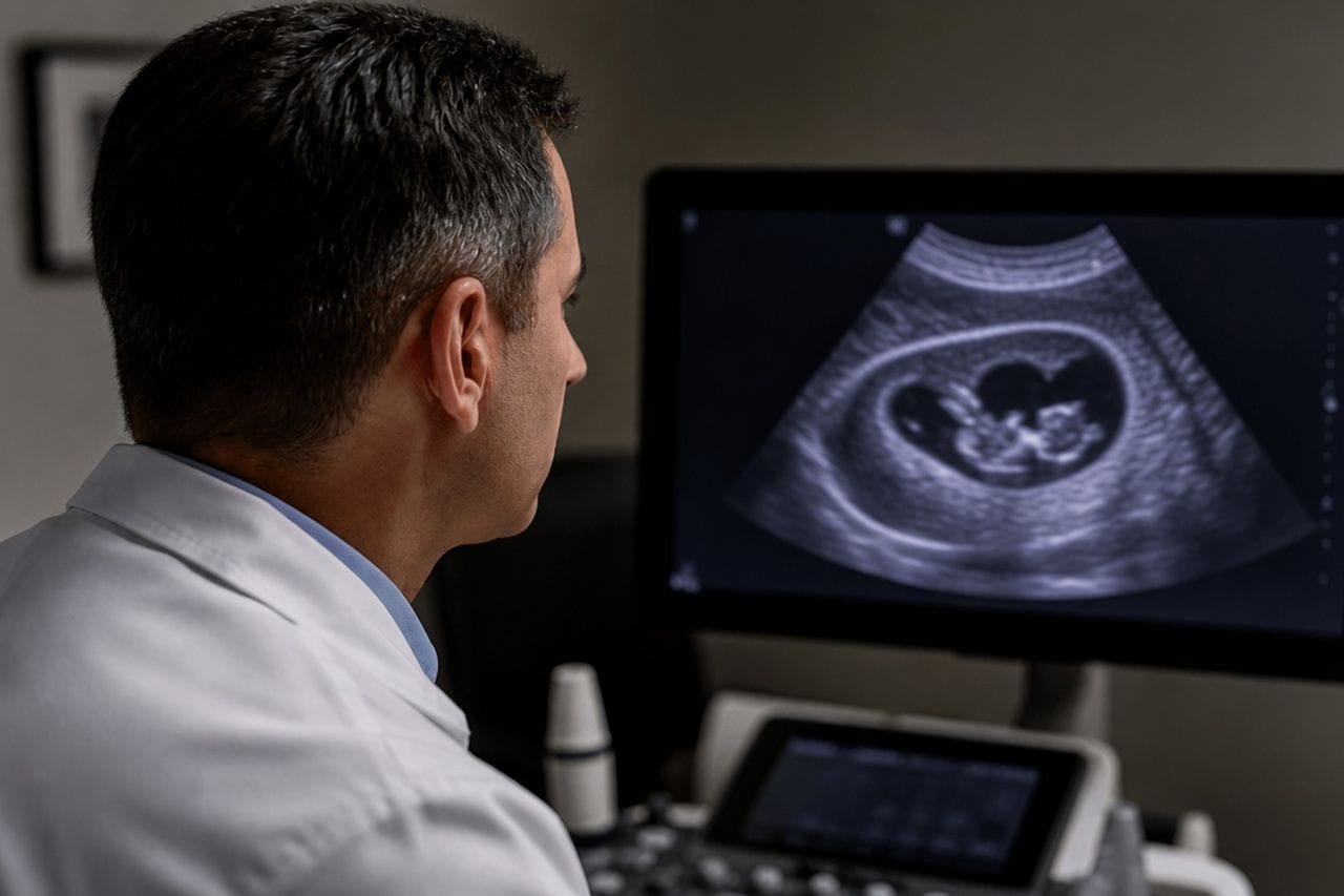
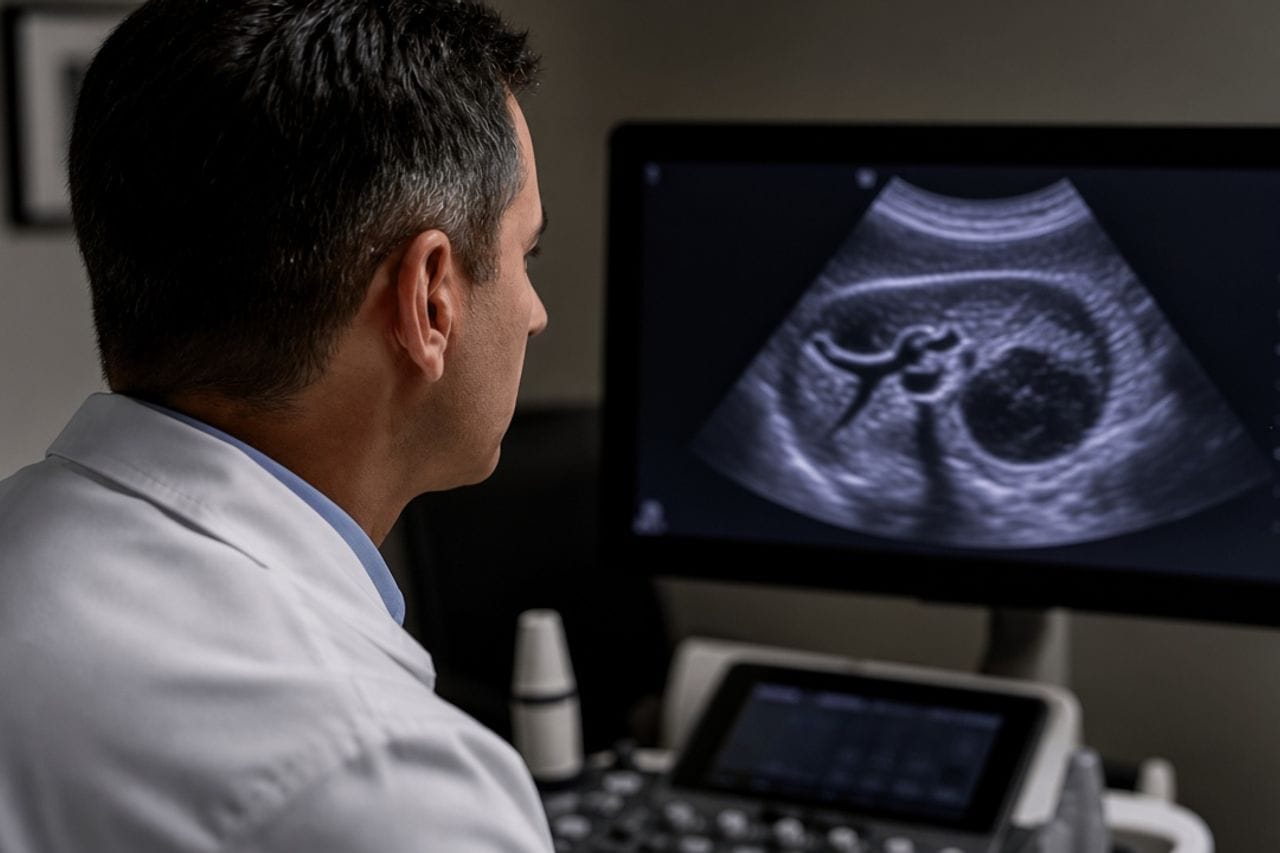
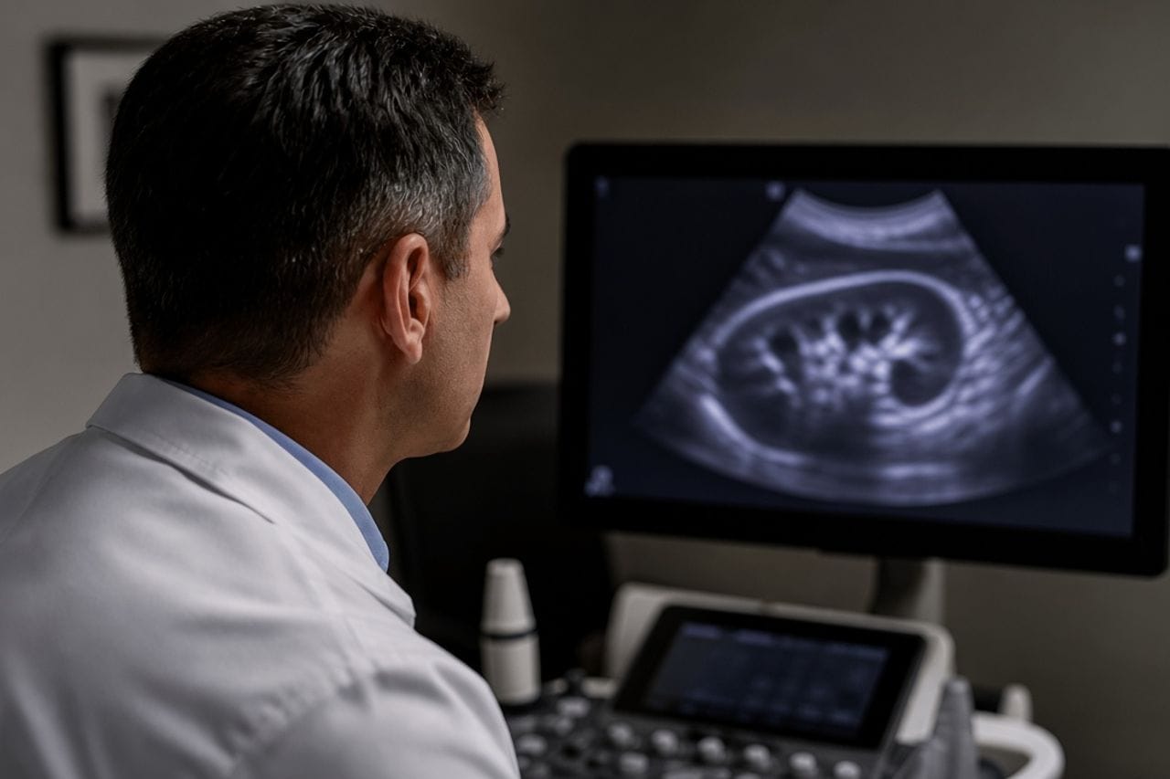
In endemic areas an echinococcus cyst is also found in the brain, more often in children and young adults. On MRI this is a rounded cavity with a thin capsule, usually without a dense node inside, as many tumors have. The fluid inside looks like CSF, and an inexperienced eye takes the cyst for "just a cyst" without a parasite.
The skull does not stretch. Even a medium-sized cyst presses on the brain, and waiting by the scheme of hepatic CE1 quickly becomes dangerous here. Neurosurgeons in such cases try to remove the cyst entirely, without opening the wall in the wound.
An alveococcosis infiltrate in the head looks different and is almost always accompanied by a liver lesion of another type: brain alveococcosis. Mixing up these two scans means choosing the wrong operation.
Which symptoms
Persistent headache, morning vomiting, seizures, unsteadiness, weakness in an arm or leg, double vision, a change in behavior. In a child — falling school performance, sudden "blackouts," in small children sometimes an increase in head circumference if the sutures have not yet closed.
The pain can last for months and is treated as migraine. Vomiting without diarrhea and without fever is more alarming than the pain itself: that is how raised pressure inside the skull behaves.
There is often no fever. If the fever is high, they also think of suppuration, but for a brain echinococcus cyst this is not the most common debut. Liver complaints may be absent even if a second cyst in the liver is there.
- Rising pain with morning vomiting — not a month on painkillers.
- MRI is needed, not a skull x-ray and not ultrasound of the head.
- Seizures for the first time in life — an exam the same day.
- The liver and lungs are checked in the same pass: a cyst in the head may not be the only one.
- A child with such complaints is not written off as gadgets: echinococcosis in children.
How they distinguish it from a tumor and from alveococcosis
A tumor more often has a node, an irregular edge, and takes up contrast differently. An echinococcal cyst is a sphere with a thin wall and contents close to water, without septa or with them if there are many vesicles. Daughter cysts inside make the picture more characteristic.
Alveococcosis gives an irregular lesion, not a sphere. If the liver on CT is infiltrated and in the head there is a node without a capsule, that is not a reason to call the finding an "echinococcal cyst" and manage it as CE. PNM and CE stages are different languages: PNM, CE1–CE5.
A brain abscess usually comes with fever, different contrast uptake, and a different history. The final word is with the neuroradiologist and the clinic, not with one phrase in a discharge summary.
Why surgery is more common here
A drug course that is sometimes started for a small active liver cyst is not the starting tactic for the head. While the cyst stands inside the skull, it occupies the place of the brain. Rupture of the cyst into the ventricles or under the membranes is a severe complication; they do not wait for it "on tablets out of interest."
The operation is built differently from liver capsulectomy. They try to take the cyst out entirely so contents do not enter the wound. If the wall tears on the table, the risk of recurrence at the operation site rises. So the words "we will just drain it" are especially unfortunate here.
A course may be added before or after surgery by decision of the neurosurgeon and infectious disease specialist. The article does not give a dose. The hepatic fork "possible without a scalpel" remains about the liver: surgery for a liver cyst.
Which other organs to look at
Chest and abdominal CT is needed almost always. A brain cyst can be the only one, but often it is not, and a missed lung or liver cyst surfaces in a year. Abdominal ultrasound without lung CT is little for this search.
Antibodies add, especially if the MRI is atypical. A negative test with a classic round cyst in a child from an endemic area does not lift the diagnosis. How serology is built: blood test.
Family screening is appropriate after the acute question with the head is resolved, not instead of MRI: family screening.
After removal
Follow-up MRI is needed on schedule, even if the head is "like new." Recurrence in the cyst bed and a new cyst in another organ are different events; both are looked at. The general logic of return after liver surgery is not copied one to one, but the habit of dropping follow-up is the same danger: recurrence.
Seizures may remain for a time after removal because of the fact of the cyst and the operation themselves. Anticonvulsants are not stopped independently in the first quiet week.
Fever, new weakness in a limb, repeat vomiting are not "that is how it heals," but a reason to contact the neurosurgeon who operated.
- Keep the operation protocol: whether the wall was opened.
- Bring the preoperative MRI to any follow-up visit.
- Ask when the next MRI is, and do not move it because you feel well.
- Check whether CT of the liver and lungs was done before discharge.
- Do not puncture any new "cyst" in the abdomen without regard to the head history.
Review at Visus Medical
Send the MRI. We say whether the image looks like a parasitic cyst, how it is not alveococcosis, and what to look at besides the head, before you are taken to surgery "for a tumor."
The head with clear pressure is managed by a neurosurgeon, and we do not replace that with a promise of a course. Our part is not to miss a second site and not to mix up the type of parasite.
Echinococcosis. The MRI disc and abdominal scans are better carried in one folder.
Need an echinococcosis consult?
Message us on Telegram or WhatsApp — we’ll review your scans and suggest a plan.