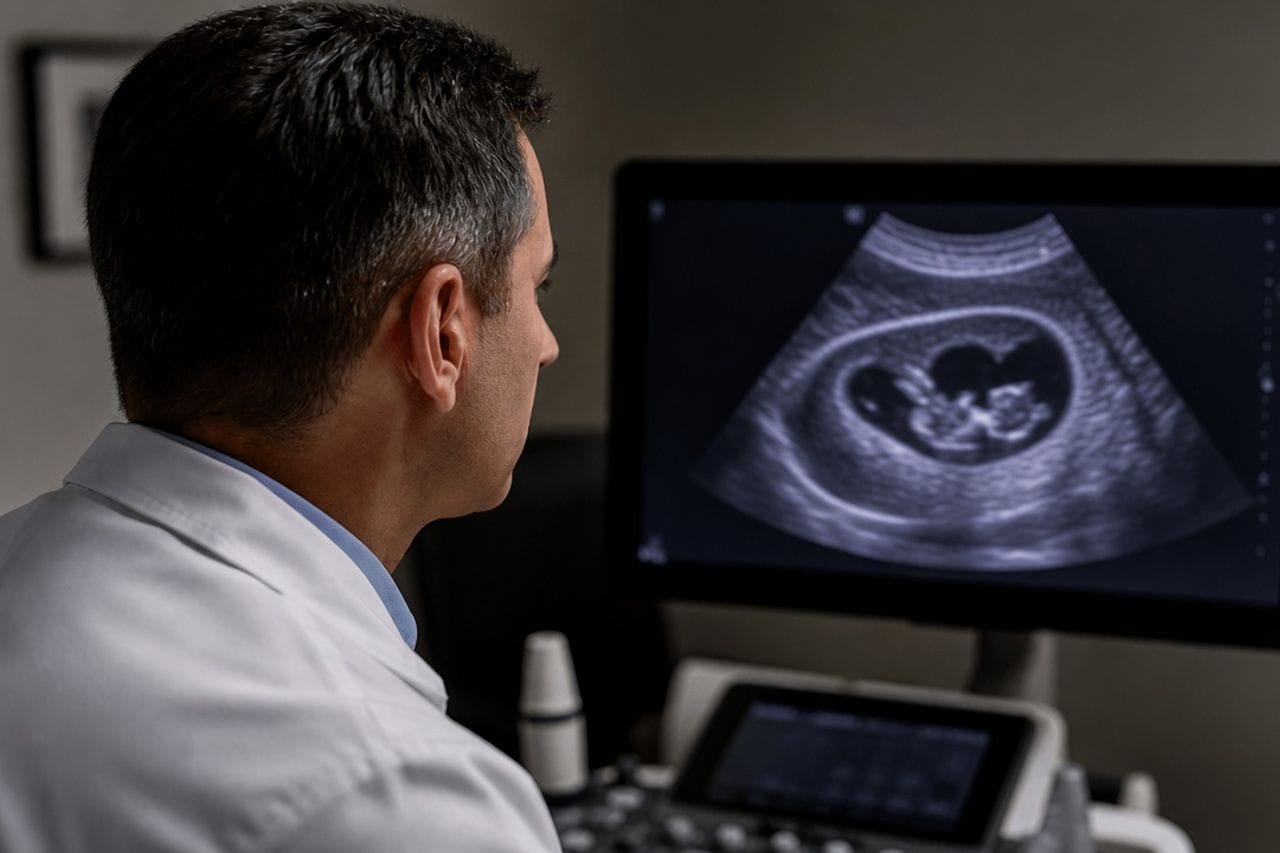
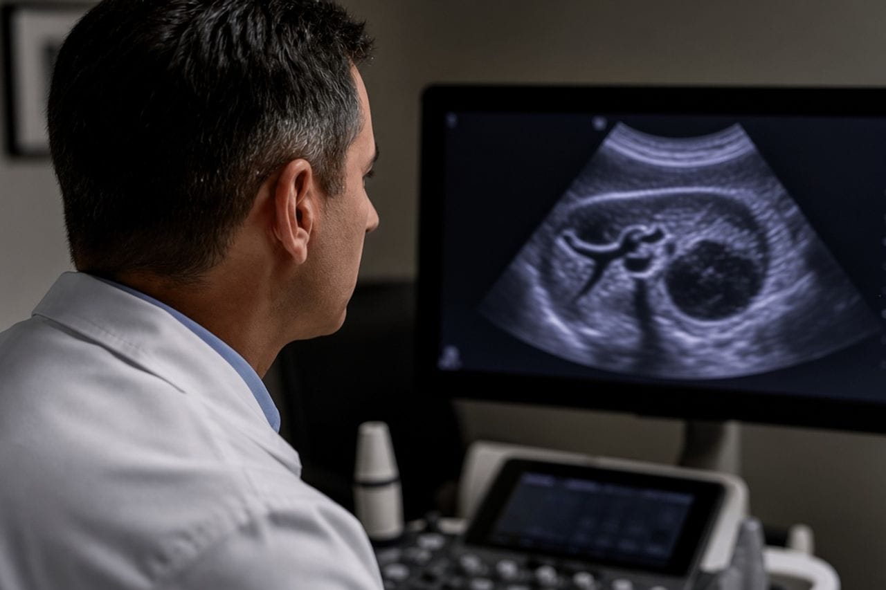
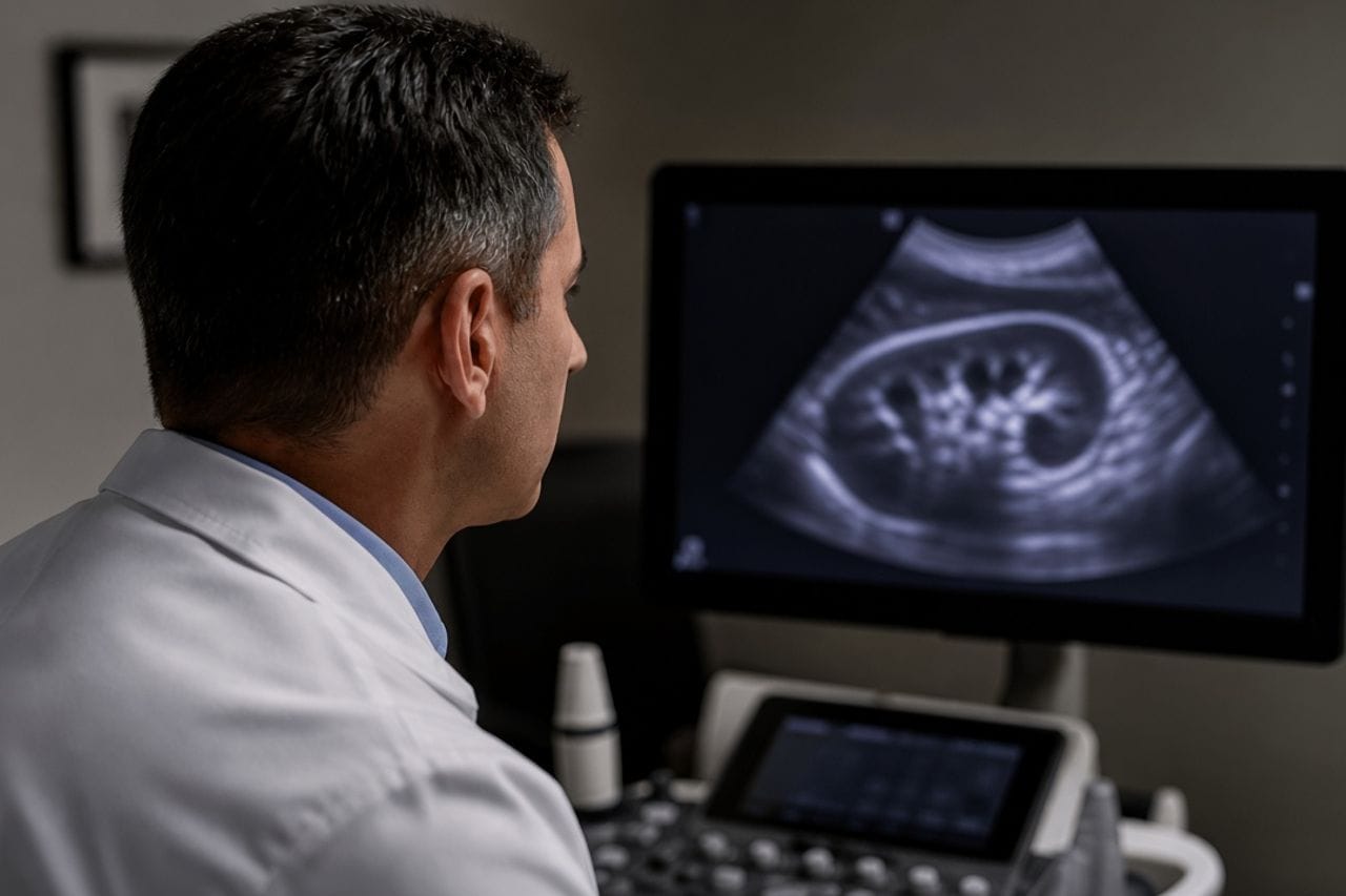
The primary echinococcus cyst more often sits in the liver, less often in the lung. If it opened into the abdomen, daughter elements settle on the peritoneum, omentum, mesentery, sometimes near the pelvis. Months and years later they already find a scatter, although the person remembers only one cyst or one attack of pain.
This is secondary abdominal echinococcosis. It is not the same as "many cysts inside the liver itself," although the words on the form look alike. In the liver each cyst lies in the organ tissue. On the peritoneum they lie between organs, and surgery is different because of that.
How the moment of rupture itself looks and why it cannot be waited out at home: cyst rupture. Several lesions in different organs without a rupture history: multiple cysts.
Where the scatter comes from
One tear is enough. Part of the contents leaves the same day and gives pain and a rash. Part goes unnoticed, and new cysts grow silently. The person arrives already with "multiple involvement" and does not link it to colic from two years ago.
A second source is an operation in which the cyst was opened in the wound and contents entered the abdomen. So in old discharge summaries the wording about how the cyst was removed and whether there was a spill matters. Recurrence next to the liver and cysts over the whole peritoneum are related but not the same stories: after surgery.
Less often a primary peritoneal cyst is described without a clear past rupture. For the patient the practical conclusion is one: count lesions over the whole cavity, and do not argue about the word "primary" until there is a full CT.
Which scans actually count the cysts
Abdominal ultrasound is convenient for a large liver cyst and poor at counting small lesions on the peritoneum, especially if they hide behind bowel. For a map you need CT of the whole abdominal cavity, not a "liver cyst close-up" frame.
The chest is imaged in the same pass. Lung and liver in echinococcus often go together, and after rupture into the abdomen a lung cyst may still have been there from the start.
Serology adds to the picture, but a scatter on CT is not cancelled by a negative ELISA. And the reverse: a plus on the test without a single lesion on full CT is not yet a diagnosis of abdominal echinococcosis. How to read antibodies: blood test.
- Ask for CT of the whole abdomen, not only ultrasound of the right upper quadrant.
- Ask whether the cysts are in liver tissue or already on the peritoneum and omentum.
- Bring the discharge about an old operation or a night of sudden pain.
- Small lesions are not the same as a task to open the abdomen and remove all of them.
- A repeat CT is compared with the previous one, not described again blindly.
Which complaints happen, and which do not
Many live without symptoms until a cyst becomes large or compresses bowel. Then heaviness, an enlarging abdomen, episodes of bloating, and less often a picture of obstruction appear. Fever hints at suppuration of one of the cysts, not at "all of them at once."
Pelvic pain in women is sometimes blamed on gynecology, although the cyst sits in the pelvis secondarily. This is not a reason for an independent puncture on the exam chair.
Absence of pain does not mean there are few lesions. An asymptomatic scatter is typical, so the scan matters more than how you feel.
Why they do not cut every cyst
One large cyst with pain, bowel block, or doubt whether it is a tumor is a reason to call a surgeon. Dozens of small ones on the omentum often cannot be removed in one operation without heavy trauma, and after such an intervention new lesions are still possible if the spill repeats.
So with secondary peritoneal involvement they more often discuss a course that acts on the parasite at different points, plus a targeted operation where there is a complication. This is not a promise that "tablets will remove all cysts by next month." Duration and the drug are chosen by the doctor; there are no doses in the text.
The logic for one cyst inside the liver is different; it is not copied onto the peritoneum automatically: surgery for a liver cyst.
How not to confuse it with other diseases
Peritoneal tuberculosis, carcinomatosis, ordinary ovarian cysts, and lymphoceles after operations look similar on individual frames. The combination decides: a round capsule, daughter elements, calcification, a liver cyst in the history — not the single word "mass."
Alveococcosis on the peritoneum looks like infiltrate and nodes, not like a set of bubbles. If the borders are ragged, that is a different review: this is not a cyst.
A "just in case" biopsy through the abdominal wall when echinococcus is suspected is not ordered first. A puncture can seed the needle track.
- Do not agree to laparoscopy "just to look" until there is a CT map.
- Ask whether tuberculosis and a tumor process were excluded if the picture is atypical.
- A cyst in the pelvis is not automatically a gynecological operation.
- After any intervention you need a follow-up plan, not one discharge summary.
- The family is worth checking separately: screening.
Review at Visus Medical
Send CT of the whole abdomen and, if you have them, old descriptions of an operation or a night of sudden pain. We sort out what lies in the liver, what is on the peritoneum, where a surgeon is needed, and where a course and follow-up are more reasonable.
If they offer to "remove everything in one go," a second reading of the scans often changes the volume of surgery. Small quiet lesions and one complicated cyst are not one tactic.
Echinococcosis. The disc is needed in full: on a film of three frames the scatter is not visible.
Need an echinococcosis consult?
Message us on Telegram or WhatsApp — we’ll review your scans and suggest a plan.