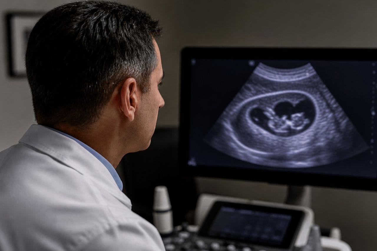
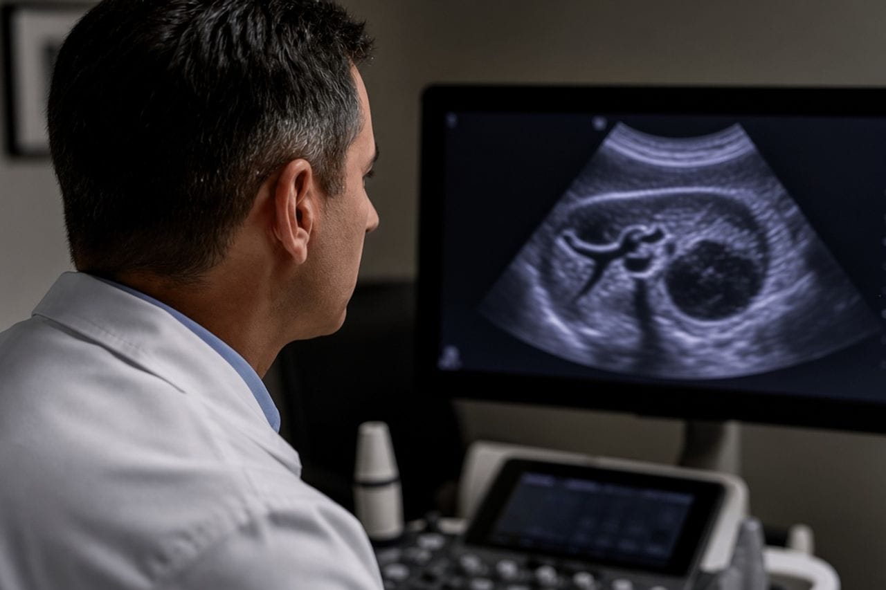
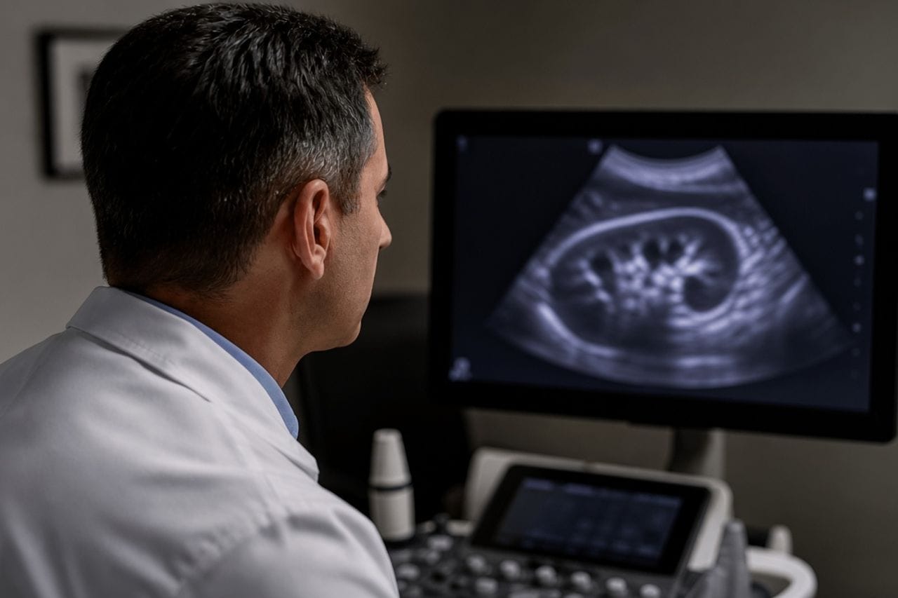
Bone echinococcus is rare, but the spine is a typical place for it, next to the pelvis and ribs. The parasite goes inside the bone, the vertebra loses strength, and a soft-tissue component may grow nearby in the canal. For years this is called osteochondrosis until the legs weaken.
On x-ray a hole in the bone is seen late. An intervertebral disc hernia looks different and does not destroy the vertebral body. If the description has "destruction," "lesion in the vertebral body," "volume in the canal," hernia ointment does not close that.
Cysts in the liver may be present or may be absent. A bone lesion is not cancelled by the phrase "abdominal ultrasound is clear."
Which symptoms
Local back pain that does not go away at rest and poorly obeys ordinary "radiculitis" schemes. Night pain is a common story. Later numbness, weakness in the legs, unsteadiness, girdle pain.
A threatening set is weakness in the legs building over days, numbness of the perineum, retention or incontinence of urine. This is compression of the spinal cord or roots, and this is an ambulance, not a visit to a manual therapist.
Fever happens with suppuration of the soft-tissue part and steers the thought toward tuberculosis or an abscess. Both versions still require scans, not an antibiotic blindly.
- Back pain with destruction of a vertebra on a scan is not a hernia.
- Weakness in the legs and a urinary problem — the same day.
- X-ray shows a late hole. For diagnosis you need MRI of the region, often plus bone CT.
- An open biopsy "just in case" is not done first: tissues can be seeded.
- The liver and lungs are still looked at: bone may not be the only lesion.
What it looks like on scans
The vertebral body is destroyed, the endplates suffer, in the canal there is a cystic or multiloculated component. Tuberculosis, a metastasis, and a primary bone tumor enter the same differential. The radiologist is obliged to name them. A parasitic version is obliged to be voiced if the person is from an endemic region or already had echinococcus.
MRI better shows the canal and soft tissues. CT better shows how much bone is eaten. Often both are needed. PET does not replace this set if the question is whether the spine can be stabilized.
Bone alveococcosis happens, but more rarely, and usually with an already known severe process in the liver. An irregular infiltrate without vesicles is read separately from classic cystic destruction: alveococcosis and cancer.
Why biopsy is dangerous as a first step
A needle or an open scrape through parasitic tissue carries contents into the muscles and the wound track. Then recurrence sits not only in the vertebra. If the scan is typical and the liver confirms echinococcus, the diagnosis is often assembled without "just looking with a knife."
When histology cannot be avoided to distinguish a tumor, biopsy is planned where they are ready for parasitic contents, not in an outpatient dressing room. This is a consilium decision.
Ultrasound puncture of a "cyst by the spine" in a clinic office is exactly the step this article warns against.
Who manages it and what to expect from treatment
With spinal cord compression neurosurgeons decide: the canal must be freed, unstable bone is sometimes fixed. This is not a scheme of observing a small liver cyst and not a promise that tablets will replace decompression if the legs are already weakening.
After the acute stage they look for remaining lesions and decide whether an antiparasitic course is needed as an addition to surgery. There are no doses in the article. Bone is a poorly accessible place; one operation without further follow-up is often not enough: recurrence in bone is known.
If there is no compression, the lesion is small, and the diagnosis is not ironclad, haste with a large approach is also not a blessing. Serial scans and a full search of other organs sometimes change the volume. A scatter in the abdomen: multiple cysts. Liver surgery is a different conversation: cyst removal.
What to bring and what not to do
MRI of the whole painful region, bone CT, abdominal ultrasound or CT, chest x-ray or CT, old back scans if the pain has lasted for years. Discharge summaries that already wrote "tuberculosis in question" or "metastasis" are also needed: they must be reread, not thrown away.
Manual therapy and traction with a destroyed vertebra can worsen compression. Heating pads and "reducing a disc" are not treatment here.
The family is worth examining after the back is safe, by the same principle as in hepatic echinococcosis: screening.
- Do not wait for weakness in the legs to "pass by itself by Monday."
- Do not start antituberculosis drugs before a reread of the scans if the diagnosis is only assumed.
- Ask whether they looked at the canal, not only the vertebral body.
- After surgery learn the date of follow-up MRI in advance.
- New urinary retention after discharge is urgent again, not a dressing in a week.
Review at Visus Medical
Send spine MRI and abdominal scans. We say whether the picture pulls toward echinococcus, how urgently to see a neurosurgeon, and whether cysts in the liver and lungs have been missed.
If you are already on a list for "removal of a vertebral tumor," a pause to reread the disc is appropriate only while there is no weakness in the legs and no urinary problem. With compression the canal comes first; an argument about the name must not cost hours.
Echinococcosis. The MRI series is needed in full: on a printed scan of two frames the canal cannot be assessed.
Need an echinococcosis consult?
Message us on Telegram or WhatsApp — we’ll review your scans and suggest a plan.