The spleen is a rare but known site of cystic echinococcosis. The report writes "splenic cyst" or "parasitic cyst." The principle is the same as for the liver: a capsule, fluid, WHO stage. This is not an alveococcosis infiltrate, although the word "mass on the left" frightens the same way.
Simple splenic cysts, aftermath of trauma, an abscess, and some tumors give a similar picture on one ultrasound. The decision is from CT or MRI and from the clinic, not from one phrase.
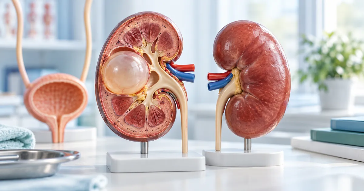
The liver is always checked. An isolated spleen happens; combination with the liver is more common. A rare kidney site for comparison: kidney.
Complaints and findings
Heaviness on the left under the rib, pain on a deep breath, a feeling of fullness after eating. Often the cyst is found by chance. A large cavity presses on the stomach and the left kidney. Fever hints at suppuration. Rupture gives sudden pain on the left and sometimes a rash: that is already an ambulance, the logic the same as for hepatic rupture: cyst rupture.
In a blood test eosinophils are sometimes raised or there is anemia if the spleen is large. This is not a diagnosis. A normal test does not cancel the cyst.
Pain on the left is blamed on the heart, the stomach, and intercostal neuralgia. If ultrasound has already shown a cyst, those versions do not cancel a scan; they go alongside.
- Do not put CT off if ultrasound says "unclear cyst on the left."
- Serology is a plus; a negative ELISA does not exclude the cyst.
- Splenectomy is not an automatic answer to the word "echinococcus."
- The liver at the same visit is mandatory.
- A spleen puncture is not done in an outpatient setting.
How they confirm it
CT or MRI shows the wall, daughter vesicles, calcification, and the relation to the splenic hilum. The hilum decides whether the organ can be preserved. Ultrasound is convenient for follow-up of an already clear cyst, and weak for the first decision.
CE stage is read by the same rules as in the liver: CE1–CE5. Active CE1–CE2 and calcified CE5 are a different conversation about the knife.
Antibodies are given, but they are not waited a week if the cyst is huge or there is fever. ELISA review: blood test.
When the spleen is removed
Organ removal is discussed for a giant cyst that replaces the spleen, for rupture, for suppuration that cannot be solved otherwise, for a suspicion of a tumor that has not been lifted. After splenectomy a person lives without a spleen, and that is a separate conversation about infections; it cannot be hidden behind the phrase "the organ is not vital."
Preserving the spleen is possible when the cyst is peripheral and can be removed with the capsule without spilling contents. This is surgery of a specific case, not a promise of an article.
An active cyst of moderate size without complications is sometimes managed with a course and follow-up, like some liver cysts. There are no doses. If there are many cysts and also in the liver, one splenectomy does not close the problem: multiple cysts.
How this is not alveococcosis and not lymphoma
Alveococcosis gives an infiltrate, more often from the liver into neighboring tissues, not a spherical capsule of the spleen. If the edge is ragged and the liver is already "like a tumor," read another text: this is not a cyst.
Lymphoma and a cyst are different densities and a different clinic. An enlarged spleen without a cavity is not equal to echinococcus. Do not agree to removal "just in case" until CT has separated these versions, if there is no rupture and a purulent abdomen.
A traumatic cyst after a blow has its own history. It is not treated with an antiparasitic course only because of size.
After the decision
If the organ was removed, you need a vaccination and follow-up plan the surgeon gives, plus liver follow-up for echinococcus. If the cyst was removed in part or a course was chosen, follow-up ultrasound or CT is compared with the baseline disc. Appearance of a new cavity on the left is not "fluid after surgery" until that is said from the scan.
Recurrence next to the splenic bed happens if contents spilled. The general principle: recurrence after surgery.
Fever and sudden pain on the left after discharge — contact the operating doctor, not waiting for a planned ultrasound in a quarter.
- Ask before surgery whether the organ can be preserved and why not.
- Clarify CE stage, not only "an 8 cm cyst."
- Take the disc and the protocol.
- Do not drop a course after splenectomy without asking: the liver may still be in play.
- The family is checked separately: screening.
Further reading
Review at Visus Medical
Send abdominal scans. We say whether this is a parasitic cyst, whether splenectomy is needed or there is a path without organ removal, and whether the main problem sits in the liver.
A referral to "remove the spleen" from one ultrasound is exactly a reason to show the disc before admission, if there is no rupture and high fever with pain.
Echinococcosis. Full CT is more useful than a frame of only the left upper abdomen.
Discuss your findings and care options
Need an echinococcosis consult?
Message us on Telegram or WhatsApp — we’ll review your scans and suggest a plan.