The kidney is not the most common site of cystic echinococcosis, but the report "kidney cyst, a parasitic one cannot be excluded" occurs. The parasite is the same as in the liver: Echinococcus granulosus, a cavity with a capsule. Alveococcosis in the kidney is rare and usually as a neighbor of an already large hepatic lesion, not as a separate "kidney cyst."
Not every kidney cyst is echinococcus. Simple cysts in adults are found constantly; they are thin-walled and quiet. Hydronephrosis, an abscess, a tumor with breakdown look different, but on a short ultrasound form they can all stand in one sentence.
So tactics start with a proper scan, not with the phrase "remove the kidney at once." The liver with any suspicious kidney cyst is looked at without fail: the combination is more common than an isolated kidney.
Which symptoms
For a long time there are no complaints. The cyst is found on ultrasound "for blood pressure" or for low-back pain that is actually from the back. Later — heaviness in the side, dull pain, blood in the urine, fever if the cavity has suppurated. Colic happens when the cyst or its contents interfere with outflow from the pelvis.
Blood in the urine does not prove a parasite and does not cancel it. Stones, a tumor, and infection stay on the list. You cannot write blood off as "sand" with an already known thick-walled cyst.
Sudden abdominal pain and a rash are not a typical debut of a kidney cyst, but a reason to recall rupture of a hepatic one if it was there too: rupture.
- First they separate a simple cyst and a tumor; they do not set echinococcus from one word.
- In parallel they image the liver and, if possible, the chest.
- A biopsy of a kidney cyst is as a rule not done: a puncture seeds.
- Blood in the urine and fever do not wait a planned month.
- A negative ELISA does not cancel a thick-walled cyst.
What ultrasound shows and why CT
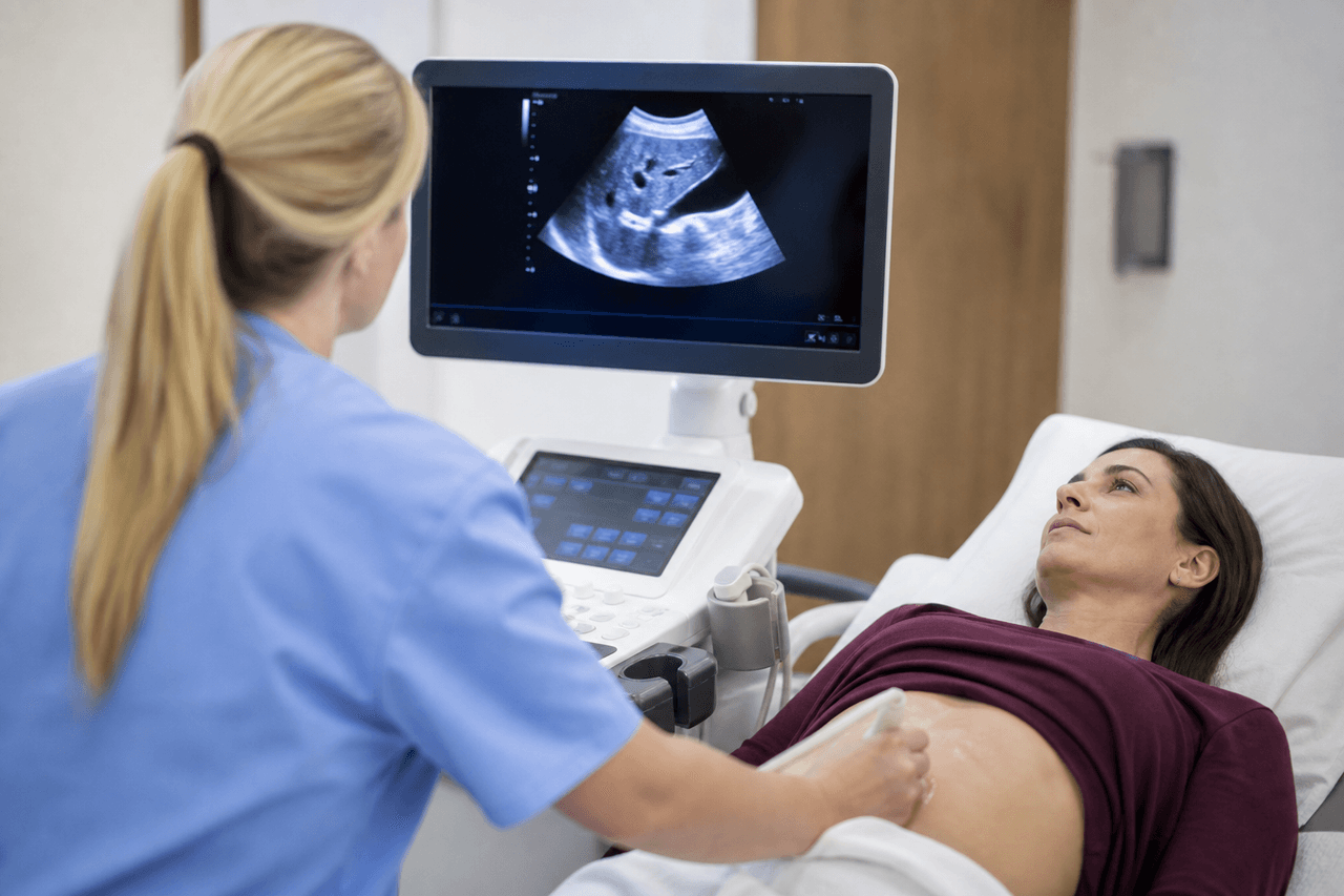
A thick layered wall, septa, daughter vesicles, and calcification in the wall are suspicious. A simple cyst is a thin wall and homogeneous water. Ultrasound hints at this. CT or MRI shows where the cyst is relative to the pelvis and vessels and whether there are the same lesions in the liver.
They try to set CE stage the same way as in the liver. It decides whether the cavity is alive. The scale: CE1–CE5. Suppuration masks the stage and adds fever: suppuration.
Antibodies are a plus to the scan. How not to overvalue them: blood test.
When kidney removal is discussed
The kidney is removed entirely if the cyst has destroyed it, the pelvis is dead, there is a complication that cannot be solved with organ-sparing surgery, or the diagnosis swings toward a tumor and the kidney can no longer be saved. This is not the default answer to the word "echinococcus."
Organ-sparing removal of the cyst with the capsule is possible with a suitable position and stage, in the hands of a urologist who can keep from opening the cavity in the wound. A blind "we will open and drain" for a parasitic cyst is a risk of seeding the retroperitoneal fat.
A small active cyst without pelvic block is sometimes managed with an antiparasitic course and follow-up, by the same logic as some hepatic CE. There are no doses in the article. The decision is only from scans. The hepatic fork for comparison: surgery or not.
Kidney plus liver
If there are cysts in both organs, kidney removal does not treat the liver and vice versa. A map of lesions matters more than a heroic operation at one address. Multiple involvement: several cysts.
The spleen is nearby in rarity, but it is another organ and another operation: spleen.
After any kidney operation follow-up is needed both of the low back and of the liver. Recurrence in the bed and a new cyst in the abdomen do not exclude each other: after surgery.
What not to do
Do not puncture a kidney cyst in a clinic "to learn the composition." Do not start a course at a dose from the internet before CT: you can treat a simple cyst in vain or be late with a pelvic block. Do not heat the low back with fever.
Do not agree to kidney removal on the day of the first ultrasound if there is no fever, blood that will not stop, and purulent destruction. There are usually a day for CT and a second reading. If the kidney already does not work and there is sepsis, those days are not stretched into a month.
The family is worth looking at after the plan for the kidney is clear: screening.
- Bring ultrasound of the kidneys and liver, not only the text about the kidney.
- Ask whether the pelvis is alive and whether there is outflow.
- Clarify the stage, not only centimeters.
- Take the CT disc if contrast was already done.
- Blood in the urine after discharge — to the urologist again, not "that happens for a week" without contact.
Further reading
Review at Visus Medical
Send ultrasound and CT of the kidneys and abdomen. We say whether the cyst looks like echinococcus, whether a course without organ removal can be discussed, and whether lesions in the liver have been missed.
If kidney removal is already scheduled, the disc is still worth showing while there is no emergency purulent destruction. Some of these referrals after CT narrow to cyst removal or to a course.
Echinococcosis. An ultrasound report without a picture is especially little for the kidney: the wall and septa are simplified in the text.
Discuss your findings and care options
Need an echinococcosis consult?
Message us on Telegram or WhatsApp — we’ll review your scans and suggest a plan.